Bacterial Pneumonia
Pasteurella and Mannheimia Pneumonia
Pneumonic pasteurellosis is a Cranioventral fibrinous bronchopneumonia. The disease occurs in goats throughout the world.
Etiology
Both Pasteurella multocida and Mannheimia (previously Pasteurella; Angen et al.
1999) haemolytica cause pneumonia in goats (Ojo 1977). Both species are Gram-negative, tiny, ovoid rods that do not form spores. They grow well on blood agar, where only M. haemolytica causes hemolysis. Colonies are 1-2 mm in diameter. Pasteurella multocida but not M. haemolytica produces indole.Mannheimia (Pasteurella) haemolytica was previous divided into two biovars: biovar A, which ferments arabinose, and biovar T, which ferments trehalose (Bingham et al. 1990). Subsequently, the T biovar was designated as Pasteurella trehalosi, and then the organism was assigned to a new genus, becoming Bibersteinia trehalosi (Blackall et al. 2007). How this might clarify the epidemiology of pasteurellosis in goats remains to be determined. The trehalosi organism has been cultured from the pharynx of healthy pack goats (Ward et al. 2002). A biotype T isolate obtained from an acute fatal case of caprine pneumonia has been used to experimentally produce a proliferative and exudative pneumonia (Ngatia et al. 1986). Biotype T M. haemolytica has also been cultured in association with an outbreak of CCPP in Ethiopia (Shiferaw et al. 2006). When typing has been performed of Mannheimia isolates from goats with pneumonia, M. haemolytica type A2 has been reported most frequently (Fodor et al. 1984; Midwinter et al. 1986; Hayashidani et al. 1988).
Epidemiology and Pathogenesis
All of these organisms commonly reside in the upper respiratory tract of normal goats. While a previous viral infection would increase the probability of lung invasion by Pasteurella or Mannheimia, field conditions often provide enough stress for the organism to be a primary pathogen.
Poor ventilation is a major factor permitting invasion of the lungs, but crowding, parasitism, and malnutrition all contribute to development of disease (Brogden et al. 1998). Some affected goats have been recently stressed by transport (Mugera and Kramer 1967), and steroid administration appears to increase the proliferation of M. haemolytica in the nasal passages of transport-stressed goats (Jasni et al. 1991).The virulence of these organisms is either very high or increases during an outbreak, so that the disease may spread to unstressed herd members (Pande 1943). In other instances, pneumonia is recognized only in animals exposed to newly introduced goats (Hayashidani et al. 1988; Buddle et al. 1990b).
A ruminant-specific leukotoxin produced by M. haemo- lytica is believed to be very important in the pathogenesis of pasteurellosis (Shewen and Wilkie 1985; Zecchinon et al. 2005). This toxin impairs and lyses alveolar macrophages and neutrophils that arrive in the lung to fight the infection. Enzymes released by the dying neutrophils cause additional injury to lung tissue. The deleterious effects of other toxins and cell-associated products of M. haemolytica have been reviewed (Brogden et al. 1998).
Clinical Signs
In the acute case, there is typically a fever of 40-41.1 °C (104-106 °F) and also mucopurulent nasal and ocular discharge. Lethargy, anorexia, dyspnea, and a moist, painful cough are noted. Auscultation may reveal crackles, areas of consolidation (increased bronchial tones), or pleuritis (friction rub early, muffled sounds later). Ultrasonography will aid in demonstrating consolidation and pleuritis. Mortality rates may be 10% or more. Commonly, one goat is found suddenly dead before any are noticed to be ill (Borgman and Wilson 1955; Mugera and Kramer 1967).
Diagnosis and Postmortem Lesions
Diagnostic cultures may be obtained from tracheal washes or from necropsy specimens. Cultures taken from the nasal passages are not acceptable substitutes, although the common presence of potential pathogens in these samples has been the basis for excluding pack goats from areas with bighorn sheep.
Kids with acute pasteurellosis often have a septicemia, and bipolar organisms may be visible in stained blood smears (Ojo 1987). A radiographic examination documents the cranioventral distribution and rules out the presence of a penetrating metallic foreign body from the reticulum, which rarely acts to introduce infection into the thoracic cavity (see Chapter 10).At necropsy, the cranioventral lung lobes are usually affected bilaterally. There is a red to purple consolidation of these lobes, sometimes accompanied by a fibrinous pleuri- tis (Figure 9.7). Histologic changes are typical of pasteurellosis in other species and include hemorrhage, necrosis, and exudation of fibrin, edema fluid, and neutrophils or macrophages into airways.
A Pasteurella or Mannheimia infection may be secondary to an interstitial pneumonia or possibly may upregulate the CAE virus when macrophages are activated in the lung. If poor body condition suggests that a dead goat has been ill for weeks or months, the dorsal lung lobes should be palpated very carefully. If they seem firmer than normal,
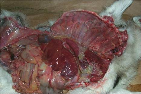
Figure 9.7 Cranial ventral localization of a Mannheimia pneumonia with marked fibrinous pleuritis. Source: Courtesy of Dr. M.C. Smith.
histologic examination to detect concurrent interstitial pneumonia should be requested.
Treatment and Prevention
Antibiotics commonly used for parenteral treatment include penicillin (20 000-40 000 IU/kg once daily), ampicillin (5-10 mg/kg twice daily), tetracycline (5 mg/kg once or twice daily), tylosin (10-20 mg/kg once or twice daily), ceftiofur (1.1-2.2 mg/kg daily), and florfenicol (40 mg/kg every one to two days SC). in vitro testing of pneumonia isolates from goats has also shown sensitivity to tulathro- mycin (Clothier et al. 2012), an antibiotic with prolonged activity from a single SC injection of 2.5 mg/kg. A sample for culture obtained by tracheal wash or at necropsy should be submitted for sensitivity testing when dealing with herd outbreaks, chronic cases, or very valuable goats.
However, caprine isolates of Mannheimia and Pasteurella are not often resistant to antibiotics routinely used for respiratory disease, as determined by the disk diffusion assay method (Berge et al. 2006). Ventilation should be corrected to decrease humidity in the barn, so that there is no condensation on windows and walls. Nutrition, including vitamin E and selenium, should be optimized. Newly introduced goats should be kept isolated from the herd for at least two weeks.In some outbreaks, young kids appear to be especially susceptible to pasteurellosis in a septicemic or pneumonic form. Colostral transfer of antibodies may be very important in preventing these infections of the neonate (Gourlay and Barber 1960).
No commercial bacterin has been proven effective against pasteurellosis in goats in the United States. For instance, vaccination with an A1 serotype bacterin did not reduce the proportion of pack goats with positive pharyngeal cultures (Ward et al. 2002). This is partly because of a multitude of serotypes possible (Ward et al. 2002) and partly because the antibodies produced in response to vaccination may contribute to damage of pulmonary tissue when infection occurs. A commercial vaccine containing multiple sheep isolates decreased lung lesions in goats in one small trial (Zamri-Saad et al. 1989). A toxoid vaccine directed against the leukotoxin produced by M. haemolyt- ica, as developed for cattle (Bechtol et al. 1991), may prove more useful, but needs further evaluation in goats (du Preez et al. 2000). A mutant strain of M. haemolytica that expresses inactive but immunogenic leukotoxin shows promise (Briggs et al. 2013). If goats become more popular models for bovine respiratory disease, an effective vaccine may eventually be developed.
Caseous LymphadenitisAbscesses in the Lungs
Corynebacterium pseudotuberculosis abscesses are most commonly found in the head and neck lymph nodes (see Chapter 3), but abscesses may form in the lung parenchyma or mediastinal lymph nodes.
In one slaughterhouse study of 25 467 goats in India, pseudotuberculosis lesions were seen primarily in the lungs of 89 (0.349%) goats, and 30 of these animals had similar lesions in bronchial or mediastinal lymph nodes (Sharma and Dwivedi 1976b).Clinical and Necropsy Signs and Diagnosis
Because the incubation period is long, this is a chronic pneumonia in adult goats and sheep. Clinical signs of dyspnea, exercise intolerance, and weight loss resemble the signs of progressive interstitial retroviral pneumonia. One to many abscesses may be present in any part of the lung. The abscesses are round, greenish yellow, encapsulated, and caseo-purulent or caseo-calcified. Radiographic studies (Grosso et al. 2018) help distinguish the pneumonia from the cranioventral, lobar distribution of pasteurellosis. Pleuritis may occur when abscesses rupture into the pleural cavity, but crackles from exudate moving in airways are not routinely auscultated. Histologic examination reveals concentric zones of necrotic neutrophils, round cells (i.e., macrophages, lymphocytes, and occasional giant cells), and fibrosis. Presence of Gram-positive diphtheroid bacteria and absence of acid-fast bacteria help to rule out tuberculosis (Sharma and Dwivedi 1976b), although both tuberculosis and caseous lymphadenitis have been reported in the same goat (Sharpe et al. 2010).
Diagnosis
In the absence of external abscesses, diagnosis is difficult without a tracheal wash or necropsy examination. If the goat is in a closed herd that has never experienced contagious abscesses, the probability of caseous lymphadenitis is minimal. Serologic test results for the organism (see Chapter 3) in affected goats are usually positive, but this cannot be used to confirm a diagnosis of the pneumonic form of caseous lymphadenitis in a herd in which goats are frequently affected with external abscesses. Positive serologic test results also occur during the incubation period of an external abscess and after its resolution by drainage.
Treatment
Long-term antibiotic treatment has little effect on the abscesses. A possible exception might be the combination of penicillin or oxytetracycline and rifampin (Chapter 3). Because the condition is most readily confused with the equally untreatable retroviral pneumonia, euthanasia is warranted when other conditions amenable to therapy have been excluded from the differential diagnosis.
Prevention
The pulmonary form of caseous lymphadenitis is reportedly common in infected flocks of sheep that are driven through a dipping vat immediately after shearing. The dipping solution becomes contaminated with pus from open abscesses and is then swallowed or inhaled by other animals in the flock. Angora and Cashmere goats should be at similar risk. Dipping should be postponed until at least two weeks after shearing if the disease is present in the flock. Another mode by which infection reaches the lungs is by drainage of the thoracic duct into the systemic circulation. Finally, aerosol transmission from another animal with pulmonary abscesses is possible, although intranasal inoculation failed to produce abscesses in experimental goats (Brown et al. 1985). Thus, culling all goats with external abscesses or chronic wasting is important to preventing pulmonic caseous lymphadenitis.
Tuberculosis
Contrary to the fervent belief of many hobbyists, goats are susceptible to tuberculosis (Ramirez et al. 2003). Goats may serve as a reservoir of infection for cattle (Napp et al. 2013) or they may directly infect humans. On the other hand, humans with tuberculosis (especially if immunocompromised by concurrent human immunodeficiency virus infection) are a potential source of tuberculosis infection for goats.
Etiology
Mycobacterium bovis typically causes pulmonary lesions in goats, whereas Mycobacterium avium is generally associated with intestinal involvement. Infection of goats has seemed to occur infrequently, even in regions where cattle and swine tuberculosis is prevalent (Nanda and Singh 1943; Thorel 1984). More recently, however, tuberculosis has been reported to be common in goats in Spain (Liebana et al. 1998). An extensive outbreak involving at least nine herds of Golden Guernsey goats in the United Kingdom was shown to be due to a cattle strain of M. bovis endemic in Wales (Daniel et al. 2009). Mycobacterium tuberculosis (human type) is a rare cause of generalized caprine tuberculosis (Sharma et al. 1985). Recently, some strains of M. tuberculosis isolated from goats, cattle, wildlife, and humans in Europe and having multiple IS6110 insertion sequences have been classified as a new species, Mycobacterium caprae (Prodinger et al. 2005). These include strains previously identified as M. bovis subsp. caprae or M. tuberculosis subsp. caprae, calling into question the species identification in the earlier literature. Mycobacterium kansasii has been cultured from the mediastinal lymph nodes of one tuberculin-positive goat during a tuberculosis eradication effort in the Canary Islands (Acosta et al. 1998).
Tubercle bacilli are Gram positive, acid fast, and aerobic. Culture is easily done in Dorset or Stonebrinks medium. Primary cultures require three to four weeks before colonies are visible. The organisms are killed by pasteurization, but may survive for a long time in moist soil or organic matter.
Clinical Signs
In countries where tuberculosis still occurs in livestock, pulmonary tuberculosis caused by M. bovis, M. caprae, and possibly other species may cause severe respiratory signs in goats or remain in a subclinical stage. Weight loss, poor milk production, anemia, and moderate coughing are possible clinical signs (Bernabe et al. 1991), but are nonspecific. Some goats also have firm nodular lesions in the udder.
Diagnosis
Diagnosis in living goats is ordinarily made by an intradermal tuberculin test, performed as for cattle. In the United States, only federal, state, and accredited veterinarians may perform a tuberculin test. A 26-gauge, 1 cm needle is used to inject 0.1 mL of PPD Bovis tuberculin intradermally in one tail fold. The test result is determined by observation and palpation at 72 (±6) hours (USDA 2006). Other countries may require injection of the tuberculin in the cervical skin, and this site is used in the United States for a comparative cervical test where the reaction to M. bovis antigen is compared to the reaction to M. avium antigen. False-positive skin tests may be seen in herds infected with or practicing vaccination against paratuberculosis (M. avium subsp. paratuberculosis), because of a potential for mycobacterial cross-reactions. Numerous dual infections with the two mycobacterial species have been observed, however, in goats in Spain (Bernabe et al. 1991). Infection with C. pseudotuberculosis is less likely to interfere with interpretation of the skin tests for tuberculosis (Bezos et al. 2015).
An appropriate serologic test may be helpful in confirming a diagnosis of tuberculosis in animals from herds that are also infected with paratuberculosis (Acosta et al. 2000). A bovine gamma interferon test for cell-mediated response using bovine PPD was positive in one tuberculin skin test-positive goat from which M. bovis was isolated, but was also positive in 12 other skin test-negative goats that were negative on culture (Cousins et al. 1993). Thus, the gamma interferon test may have poor specificity in exposed but apparently uninfected goats. Other authors propose that the gamma interferon test is more sensitive than the tuberculin test, detecting the infection at an earlier stage, and that its results should be believed during eradication programs (Liebana et al. 1998).
Various authors report caseation, calcification, and encapsulation of lesions in lymph nodes; in the parenchyma of the lung, liver, and spleen; and in the peritoneal and pleural cavities (Murray et al. 1921; Carmichael 1938; Bernabe et al. 1991; Daniel et al. 2009). When caseous granulomas are found in the lungs at slaughter or at necropsy, histology (including acid-fast stains) and culture are necessary to confirm the diagnosis. Other agents that might cause a similar pneumonia include Yersinia pseudotuberculosis (Rajagopalan and Sankaranarayanan 1944), Burkholderia (Pseudomonas) pseudomallei, Rhodococcus equi (Carrigan et al. 1988), and C. pseudotuberculosis. Dual infections with tuberculosis and caseous lymphadenitis have also been reported in goats in Ireland (Sharpe et al. 2010). Granulomatous pneumonia caused by the opportunist Cryptococcus neoformans together with M. bovis has been reported in a goat that was negative on intradermal and serologic tests for tuberculosis, suggesting an underlying immunodeficiency (Gutierrez and Garcia Marin 1999).
Control
Government regulations concerning herd quarantine and removal of infected animals must be followed. Cleaning and disinfection with Virkon® (Antec International, Sudbury, UK) or a cresol product such as 5% phenol solution should be stressed, with special attention to feed troughs and water containers. Other livestock species on the premises and the human caretakers should be tested for tuberculosis. Milk fed to goats and humans should be pasteurized. Artificial rearing and isolation have been used to preserve valuable genetics in infected herds (Liebana et al. 1998). Vaccination with the M. bovis Bacillus Calmette-Guerin (BCG) vaccine has been shown to be safe, though it would interfere with testing for the disease (de Val et al. 2016).
Melioidosis and Rhodococcal Pneumonia
Melioidosis is a disease in tropical regions caused by Burkholderia (Pseudomonas) pseudomallei. Infected goats may develop respiratory signs, including coughing and dyspnea on exercise. Abscesses often form in external lymph nodes and lungs. Abscesses are frequently 2-5 mm in diameter (but may be larger) and may coalesce in the lungs. Abscess contents are white, cream colored, or greenish, and are viscous, caseous, or dry (Thomas et al. 1988). Lung abscesses can be demonstrated radiographically (Soffler et al. 2014). Affected animals are culled because of the danger of human infection. This condition is discussed in Chapter 3.
Disseminated Rhodococcus equi infection was identified in two goats, one of which had raised nodules in the lungs containing off-white caseous material arranged in concentric layers. An enteric route of entry was suspected because of extensive abscessation of the liver. The organism isolated appeared to lack virulence antigens normally present in strains pathogenic for foals (Davis et al. 1999). Similarly, rhodococcal pneumonia in a yearling Angora was confirmed by culture of R. equi from pulmonary abscesses that ranged from 0.5 to 4 cm in diameter and resembled the lesions of caseous lymphadenitis (Fitzgerald et al. 1994). In a later report, R. equi isolates from goats with pulmonary abscesses and caseous granulomas in mediastinal and/or mesenteric nodes, as well as other organs, did carry virulence plasmids (Stranahan et al. 2018).