Clinical Anatomy and Examination of the Eye
The history elicited from the owner may suggest a problem with ocular structures or vision. The goat may have a hesitant gait, appear to be timid or hiding, or refuse to move or pass through a gate.
The animal may carry its head abnormally elevated or near the ground. A report of normal vision should be verified by the examiner; visual loss may not be apparent until the goat is placed in unfamiliar surroundings. The eyes should also be examined relative to systemic conditions (i.e., dehydration, anemia, icterus, septicemia, hemorrhagic diseases) and during prepurchase or breeding soundness examinations. A review article on clinical examination of the ruminant eye has been written by Townsend (2010).Lids and Lashes
The upper and lower eyelids normally are tightly apposed to the globe. They should neither roll inward (entropion) nor gape outward (ectropion). Lashes should not be in contact with the cornea. The examiner should note the location of any long, tactile hairs before evaluating vision by the menace response. Touching the lids near the medial and lateral canthus should evoke the palpebral reflex (blinking and retraction of the globe). This reflex requires function of cranial nerves V (sensory branches of trigeminal nerve) and VII. Conditions causing facial nerve paralysis are discussed in Chapter 5. The third eyelid (nictitating membrane) is normally unobtrusive and retracted to the edge of the medial canthus. It can be seen better if the globe is retropulsed, using digital pressure through the eyelids. Topical anesthesia permits grasping of the third eyelid with smooth forceps so that its posterior surface can be inspected for lesions or foreign bodies. Prolapse of the third eyelid occurs in some cases of tetanus. Passive prolapse of the nictitans occurs in conditions causing enophthalmos, including dehydration or emaciation, as well as in neurologic diseases accompanied by Horner's syndrome.
Meibomian gland duct openings are present on both the upper and lower lid margins (Townsend 2010).Goat Medicine, Third Edition. Mary C. Smith and David M. Sherman. © 2023 John Wiley & Sons, Inc. Published 2023 by John Wiley & Sons, Inc.
Lacrimal Glands and Ducts
The lacrimal apparatus of the goat has been reviewed by Sinha and Calhoun (1966). Lysozyme has been identified in goat tear samples (Brightman et al. 1991). Plasma cells in the nictitating gland secrete immunoglobulin (IgA) into the tear film (Schlegel et al. 2003). Mean Schirmer tear test results in normal eyes of 16 and 10-14 mm/min have been reported (Broadwater et al. 2007; Ribeiro et al. 2010a). Normally no staining or wetness occurs beneath the goat's eye, neither is there a deep recess filled with sebum rostral to the medial canthus analogous to the infraorbital gland of sheep. There are dorsal and ventral slit-like nasolacrimal punctae (Broadwater et al. 2007). The nasolacrimal duct can be cath- eterized from the ocular puncta (Moore and Whitley 1984). nystagmus (direction opposite the direction of rotation) lasts for less than 10 seconds.
Exophthalmos (protrusion of the globe from the orbit) may be due to a retrobulbar abscess or tumor and needs to be differentiated from buphthalmia (enlargement of the globe). In the latter condition, the horizontal corneal diameter of the affected eye is greater than that of the normal eye, whereas the two corneal diameters are equal in cases of exophthalmos (Townsend 2010). Orbital ultrasonographic examination or computed tomography, followed by guided aspiration of cells or fluid from the lesion, can facilitate a diagnosis of retrobulbar abscess. Enucleation (orbital exenteration) may be required to resolve this condition. Retrobulbar tumors, which include lymphosarcoma (Valentine et al. 2011) and enzootic nasal tumor, are unlikely to be cured by enucleation. See Chapter 9 for further discussion of enzootic nasal tumor.
Conjunctiva, Sclera, and Scleral Vessels
Everting the eyelids permits inspection of the conjunctiva (Figure 6.1).
This is commonly done when evaluating for anemia, which is frequently caused by severe Haemonchus infections (see Chapter 10). Rotating the head upward and to one side exposes bulbar conjunctiva and the deeper sclera and its vessels. A pale white color with almost invisible blood vessels occurs with severe anemia, and yellow sclera is indicative of icterus. A brownish discoloration of this and other mucous membranes occurs with nitrate poisoning, a purplish color with cyanotic conditions, and bright red coloration with cyanide poisoning. Vessels may appear prominent, tortuous, and enlarged (“injected”) with ocular, regional, or systemic inflammatory or toxic diseases. Cartilage and bone have been observed in the sclera of healthy goats (Tusler et al. 2017).Ocular Position
The ruminant eye normally maintains a constant position relative to the ground rather than remaining centered between the lids. This can make ocular examination difficult if the goat's head cannot be restrained in the position required to expose the structures of interest. Abnormal function of the nerves supplying the extraocular muscles (cranial nerves III, IV, VI) affects ocular position. Dorsomedial strabismus, for instance, sometimes occurs in polioencephalomalacia. Mydriasis and strabismus may occur with botulism as a result of paralysis of intrinsic and extrinsic ocular muscles (Wyman 1983). Normal nystagmus (vestibular nystagmus) occurs when the head is slowly turned to one side and has its quick phase in the same direction as the head movement. Normal postrotatory
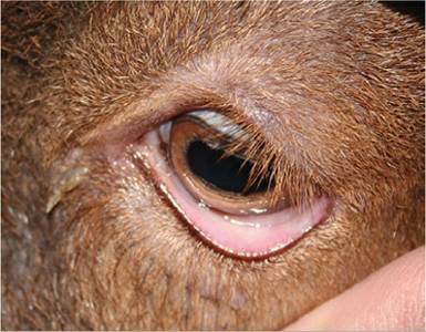
Figure 6.1 The lower lid has been everted to expose the conjunctiva. This goat had a packed cell volume of 21%. Source: Courtesy of Dr. M.C. Smith.
Cornea
The cornea is normally clear and moist. Oblique lighting and observing the clarity with which the iris can be visualized aid in differentiating cloudiness of the cornea from lens opacity.
If the cornea is lightly touched with a finger or a wisp of cotton, the corneal reflex results in blinking and globe retraction, via the same pathway as the palpebral reflex.Iris, Pupil, and Lens
A strong and well-focused light source should be used to evaluate direct and consensual pupillary response (optic nerve and parasympathetic fibers traveling with the oculomotor nerve, midbrain). Pupillary responses may be intact in a blind animal, and they may be almost impossible to elicit in an apprehensive goat. The pupil is oval, becoming more rectangular in bright light, and has granula iridica (corpora nigra) attached to both its dorsal and its ventral rim. Various toxicoses may affect the pupil, e.g., mydriasis with chlorinated hydrocarbons (Choudhury and Robinson 1950) or miosis with organophosphates. The anterior chamber, between cornea and iris, should be examined for hyphema, hypopyon, and the cloudiness characteristic of aqueous flare. Aqueous flare often indicates the presence of anterior uveitis. Anterior segment angiography using indocyanine green has been proposed as a research tool for studying the vasculature of the goat eye (LoPinto et al. 2017).
If a cataract (opacity of the lens or its capsule) is detected, the density of the cataract should be evaluated. If a portion of the retina is clearly visible in a blind animal, any cataract that may be present is not the cause of blindness.
Retina and Ophthalmoscopic Examination
The goat’s fundus can usually be examined without resorting to mydriatics. If a detailed examination is desired, and if the examiner is willing to wait 15-30 minutes for its effect, 1% tropicamide may be used to dilate the pupil (Figure 6.2). This should not be done until after pupillary response has been evaluated and diagnostic cultures or scrapings have been performed.
Hyaloid remnants on the optic nerve head and continuing into the vitreous body are commonly found in adult ruminants, including 4 of 10 goats in one study (Schebitz and Reiche 1953).
Presence of blood within the hyaloid vessel is also considered normal until 8 weeks of age in lambs, although this has not been investigated in kids.Photographs of the fundus of goats have been published (Rubin 1974; Whittaker et al. 1999; Galan et al. 2006b;
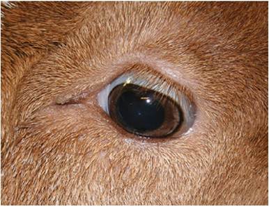
Figure 6.2 The normaLLy rectangular pupil has been dilated with tropicamide to permit a thorough fundus examination. Source: Courtesy of Dr. M.C. Smith.
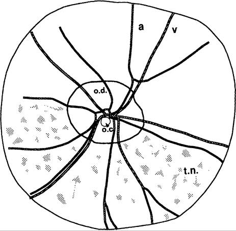
Figure 6.3 The fundus of a Saanen goat. a, arteriole; o.c., optic cup; o.d., optic disc; t.n., tapetum nigrum; v, venule.
Pearce and Moore 2013; Kalaka and Ramani 2017). A diagram of a goat fundus is provided in Figure 6.3 and a normal Boer goat fundus is illustrated in Figure 6.4. The fundus is usually in focus with a direct ophthalmoscopic setting of -1D to -5D. The optic disc is round or oval, whereas in the sheep it is often more kidney shaped. The optic disc is frequently located totally within the tapetal fundus (tapetum lucidum, yellow to bluish green). The optic disc is grayish pink and sharply demarcated, and has a small funnel-shaped physiologic cup. The broader veins enter the middle of the disc, while the thinner, redder arteries originate like rays from a pericentral location (Schmidt 1973). There are more blood vessels than in sheep
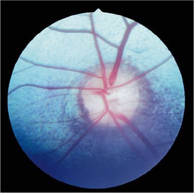
Figure 6.4 The fundus of a normal Boer goat. Source: Courtesy of Dr. M.C. Smith.
or cattle. Galan et al. (2006b) report three to six retinal arteries in the goat, often branching from a common artery that emerges from the dorsotemporal portion of the disc. There are many stars of Winslow, which are black dots in the tapetum lucidum where choriocapillaris vessels penetrate the tapetum. Fluorescein angiography of the normal goat fundus has been described (Galan et al.
2006a) using 20 mg/kg fluorescein intravenously (IV).Ultrasonography can be used to evaluate the contents of the globe and the retrobulbar area, as is done routinely in companion animal medicine. Ideally, both a 7.5 MHz probe and an offset pad are used. The ultrasound anatomy of the caprine eye using a 20 MHz probe has also been reported (Ribeiro et al. 2010b). The globe can be imaged through the upper eyelid or directly through the cornea. Tranquilization is followed by application of a local anesthetic to the cornea, and the probe is placed on lubricating jelly on the lid or on a mound of sterile ocular lubricating gel on the cornea. Normal chamber fluid is hypoechoic, while the posterior lens capsule and retina are hyperechoic. Conditions that might be demonstrated by ultrasound include hypopyon, cataract, anterior uveitis, luxated lens, retinal detachment, or a retrobulbar abscess or tumor (Toal 1996; El-Tookhy and Tharwat 2013).
including optic nerve and cerebral cortex, is also evaluated by the menace response. Fingers are moved vertically or quickly spread apart (to decrease air currents) in front of each eye in turn. Lashes and tactile hairs must not be touched. The palpebral reflex (cranial nerves V and VII) should be checked if the goat does not blink in response to the menacing gesture.