Clinical examination
Normal physical parameters and basic examination techniques are detailed by Blanshard and Bodley (2008). The signs of disease or trauma in koalas may be overt or subtle, with some clinical signs only becoming apparent late in the disease process (e.g.
cystitis, leukaemia/lym- phoma). Free-ranging koalas presented with trauma may also have underlying disease. Regular (weekly or fortnightly) weighing and body condition scoring are recommended for koalas in managed care to facilitate detection of early signs of illness. Clinical examination of any koala should also always include a distant or ‘in-enclosure’ evaluation of demeanour, posture, gait (if possible) and respiration, before detailed physical examination and/or emergency treatment. Thorough physical examination should always include the oral cavity (particularly dental health and age estimation [Blanshard and Bodley 2008]), assessment of gut fill and palpation of all superficial lymph nodes even if there are other obvious clinical signs. The use of specific diagnostic procedures (Table 33.1) are recommended for thorough assessment of any sick or injured koala.2.1 Clinical pathology
Examination of blood films, bone marrow smears, abdominocentesis smears and urinalysis (including sediment cytology) are important components in the diagnostic work-up of koalas. All can be collected safely and cost-effectively under chemical restraint, requiring relatively little experience and a basic understanding to effectively interpret findings.
2.1.1 Haematology and biochemistry
RBC morphology and biochemistry are discussed in Blanshard and Bodley (2008). Haematological and biochemical reference values are presented in Appendix 1.
Common haematological abnormalities include:
Table 33.1. Diagnostic tests indicated in addition to thorough physical examination for sick and injured koalas (Phascolarctos cinereus)
| Routine work-up for sick managed koala (Phascolarctos cinereus) | Free-ranging koala-sick managed koala work-up plus: | Severe trauma (dog attack, vehicle collision) - as for free-ranging koala plus: |
| Blood smear, TPP and PCV (for immediate assessment) and haematology and serum biochemistry USG and urinalysis Bone marrow smear Abdominocentesis Abdominal and thoracic radiography Blood gas analysis | Cystocentesis and sediment cytology* Ultrasound of UGT* Chlamydia DNA or Ag detection (ocular and UGT)* | Full-body radiography Thoracocentesis if indicated Contrast cystography and/or urethrography, if indicated |
| Additional diagnostic procedures and tests that may be indicated: exploratory laparotomy; advanced imaging (myelography, CT and/or MRI); cryptococcal antigen lateral flow assay (for screening in koalas with respiratory signs, lung opacity on radiographs or non-specific signs not explained by other signs and tests [see Chapter 25]). | ||
*Include for sick managed koala if indicated by clinical signs.
USG = urine specific gravity; UGT = urogenital tract.• stress leucogram - often seen in rescued free-ranging koalas brought into managed care and/or with chronic disease
• anaemia - acute blood loss through trauma, chronic blood loss from cystitis and/or reduced erythroid production associated with chronic inflammatory disease (poorly regenerative); seasonal blood loss associated with heavy tick and mosquito parasitism (regenerative). If blood loss is chronic and severe, iron deficiency anaemia may result, leading to a poor regenerative response
• leucocytosis - ± band neutrophils, associated with acute and/or active inflammation
• leucopenia - associated with myelodysplastic syndromes and excessive demand/depletion associated with severe infections
• free and/or phagocytosed bacteria associated with severe infections and sepsis
• vacuolated monocytes - associated with chronic inflammatory diseases, such as chlamydiosis
• leukaemia - most commonly of the lymphoid series, often with concurrent anaemia associated with myelophthisis
• mild elevations in some WBCs, including eosinophils, often without any explanatory clinical findings.
Myelodysplasia may lead to more subtle changes, such as mild to moderate anaemia, leucopenia, thrombocytopenia and sometimes large and unusually-shaped WBCs, whose lineage may be difficult to determine. A diagnosis of myelodysplasia can be supported by examination of a bone marrow smear, which may show an absence or reduction of the usual clusters of erythroid and myeloid differentiation, and in some cases a background of atypical blast cells.
Hypoproteinaemia is common in critically ill koalas and those with significant blood loss or septicaemia. Hypoproteinaemia may lead to peripheral, caecal wall or pulmonary oedema. TPP <45 g/L warrants intervention such as plasma transfusion (see section 3.1.1). Serum protein electrophoresis analyses of zoo-based and free-ranging koalas indicate that zoo-based koalas have lower albumin and β-globulin fractions as compared with free-ranging koalas, possibly as a consequence of fewer inflammatory or infectious stimuli in managed settings (Pye et al.
2012).2.1.2 Bone marrow examination
Bone marrow cytology is frequently indicated, given the relatively common occurrence of leukaemia and other bone marrow diseases, particularly in northern koalas. The technique for bone marrow sample collection is described in Blanshard and Bodley (2008). The two-slide pull-apart method should be used to prepare smears with only very gentle pressure. Three key features for assessing bone marrow cytology are as follows.
1. Overall nucleated cellularity - marked hypercellularity is associated with replacement of bone marrow with malignant or atypical cells in cases of leukaemia (Plate 33.1) and myelodysplasia (Plate 33.2). These abnormal cells are very fragile and often rupture, resulting in ‘smudge’ or ‘basket cells’. Care should be taken not to misinterpret erythroid or myeloid series responsive hyperplasia as leukaemia. Hypercellular bone marrow that is responding to increased demand shows normal differentiation and maturation of the blood cells, but large, immature blast cells may be more prominent. Myelophthisic marrow will be dominated by atypical and usually very fragile cells, with little or no evidence of normal differentiation and maturation. Hypocellularity may result from excessive suction when aspirating the sample, resulting in dilution of the sample with blood, or may be seen with hypocellular myelodysplastic syndromes such as aplastic anaemia.
2. Presence of all lineages of haemopoietic cells
- erythroid, myeloid and megakaryocytic cell lines should be easily identifiable in a normal bone marrow smear (Plate 33.3). Erythroid-lineage immature cells are nucleated and tend to have blue-purple cytoplasm and large nuclei. Normal clusters of erythroid differentiation also often contain mature erythroid cells that appear as nucleated RBCs. Myeloid series cells have more abundant and pinker cytoplasm, and clusters of normal differentiation often have cells resembling the granulocytic cell lines. Megakaryocytes are clearly visible in normal bone marrow as large cells with multinucleated or multilobulated nuclei that are less common but easily identified in smears.
3. Orderly differentiation and maturation of erythroid and myeloid series cells - correctly prepared smears of normal marrow should demonstrate clearly identifiable clusters of erythroid and myeloid differentiation (Plate 33.4). These clusters often feature a group of cells at varying stages of maturation from relatively undifferentiated blast cells through to easily identified mature cells of the erythroid or granulocyte lineage. In contrast, myelodysplastic marrow demonstrates no or poor differentiation and maturation, a background of atypical undifferentiated blast cells or simply an absence of normal haematopoietic cells (aplastic anaemia).
2.1.3 Abdominocentesis
Abdominocentesis and cytological examination of the fluid is an important component in the work-up of sick
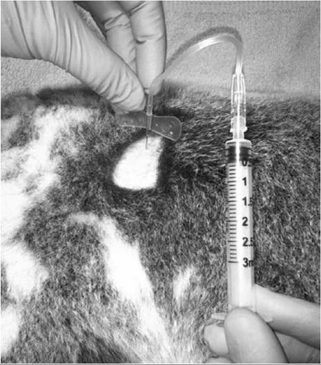
Fig. 33.1. Abdominocentesis. A 25G, 25-mm winged infusion set with a 3-mL syringe attached inserted through the skin at 45-90° to approximately one-third of the depth of the needle. Negative pressure is gently applied and the needle withdrawn to obtain a peritoneal fluid sample.
and injured koalas. An alternative method for abdomi- nocentesis to that described by Blanshard and Bodley (2008) is as follows: the sedated or anaesthetised koala is positioned in lateral recumbency (either side) and the fur in the upper region of the paralumbar fossa is parted or shaved before aseptic preparation of the site. A 25G, 25-mm winged infusion set or standard hypodermic needle with a 3 mL syringe attached is inserted through the skin (at 45-90°) to approximately one-third of the depth of the needle. Negative pressure is gently applied and the needle withdrawn (Fig. 33.1). Elevating the pelvis can aid sample collection. Normal peritoneal fluid is clear and low volume. Discolouration and/or turbidity or volumes greater than 0.25 mL are abnormal. Contamination with, or aspiration of, caeco-colic content is common, particularly if the needle is advanced too deeply.
The sample is gently smeared onto a slide (using the two-slide pull-apart technique), airdried and stained before cytological examination. Table 33.2 describes expected peritoneal fluid cytology findings for different conditions in koalas and suggested actions.2.1.4 Cystocentesis
Ultrasound-guided Cystocentesis is performed using a 3-mL syringe and 25G, 38-mm needle in an anaesthetised koala placed in dorsal recumbency. The ultrasound probe is placed just cranial to the pubis in a sagittal or transverse plane. The needle is advanced between the probe head and the pubis into the bladder, ensuring that it is maintained within the plane of the sonographic image. On entering the bladder, a 2-mL sample is aspirated and the suction stopped before withdrawing the needle. Allowing suction to continue during withdrawal of the needle can result in aspiration of peritoneal and hindgut contents, confusing the cytological findings. Urine should be centrifuged and the sediment pellet collected and smeared on a glass slide with a small-tipped swab (such as the Copan® 160C rayon-tipped sterile dry swab, Copan Diagnostics Inc., CA, USA) before drying and staining for microscopic examination.
Normal koala urine may contain scant urinary crystals and spermatozoa in males. The presence of neutrophils in urine sediment (collected by cystocentesis only) confirms cystitis and is most commonly associated with chlamydial infection. Voided or samples collected via catheter may contain neutrophils derived from other sites, such as the urethra, prostate and urogenital sinus. The presence of RBCs suggests bladder mucosal ulceration, fungal/candidal elements confirm fungal cystitis (Plates 33.12 and 33.13), whereas bacteria (Plate 33.14), urinary casts and urinary crystals (Plates 33.14 and 33.15) suggests non-chlamydial (or complicated chlamydial) cystitis or renal disease such as oxalate nephrosis.
3.