Devocalization
Certain goats bleat almost continuously when separated from something they want, such as human companionship, or during estrus. Providing the goat with a pen mate (another goat or sheep, a cat, or even a pony) may alleviate the problem.
Ovariectomy (see Chapter 13) may solve the problem of vocalization during estrus. In other cases the goat persists in its vocalizations, and the family, the neighbors, or the research group threaten mayhem. Displeased neighbors are especially dangerous to suburban goats because most zoning laws classify the pet wether as livestock. Under these circumstances, surgical removal of the vocal cords might be justified, but it would be better to find a new home for the goat.Two techniques of devocalization are described in the literature and summarized here. Anesthetic agents that might be used include xylazine/ketamine and barbiturates. These are discussed in detail in Chapter 17. Atropine (0.08 mg/kg) administered intramuscularly 20 minutes before induction of anesthesia is recommended to decrease salivation (Tillman and Brooks 1983).
Traditional Surgical Technique
For the older surgical approach (Durant 1974), the goat is positioned in dorsal recumbency with the head lower than the neck. This is achieved by placing a sand bag beneath the neck. Food should be withheld for 24 hours and water for 6 hours before surgery; it is not possible to use a cuffed endotracheal tube to prevent regurgitation and aspiration during the surgery. Skin over the larynx is prepared as for an equine roarer operation. A midline skin incision 35-40 mm long is made, with careful attention to hemostasis. The first tracheal ring is incised and that incision is carried forward on the midline through the cricoid cartilage and crico-thyroid ligament to but not through the anterior border of the larynx (see Figure 9.10a). A small divider is used to spread the larynx and expose the vocal cords.
Each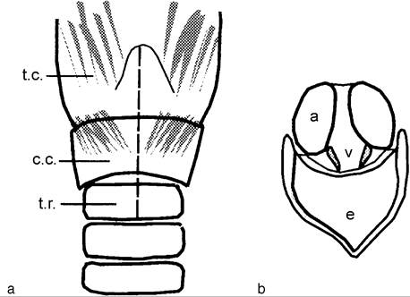
Figure 9.10 Removal of the vocal folds for devocalization. (a) Ventral view of larynx showing initial incision through cartilages of larynx. (b) Larynx as viewed from oral cavity. a, arytenoid cartilage; c.c., cricoid cartilage; e, epiglottis; t.c., thyroid cartilage; t.r., first tracheal ring; v, vocal folds.
cord (about 6 mm long) is clamped and removed down to the surface of the cartilage with curved scissors. After hemostasis is achieved, the divider is removed and the skin incision closed.
Electrosurgical Technique
The second technique involves electrosurgical destruction of the vocal cords via the oral cavity (Tillman and Brooks 1983). The anesthetized (ketamine/xylazine) goat is placed in sternal recumbency with its chest on the ground plate of an electrocautery unit. A 38 cm tonsil snare arm (without the snare wires) is allowed to protrude 2-3 mm from its insulating sheath and is used as a long electrode. A long laryngoscope blade for ruminants (Soper laryngoscope blade 257 or 385 mm, Penlon, Abingdon, UK) is also needed. The goat’s head and neck are fully extended by an assistant and the tip of the laryngoscope blade is used to depress the epiglottis. The vocal cords are positioned just posterior to the arytenoid cartilage. They are much smaller than the arytenoids and almost meet ventrally, forming a “V” (Figure 9.10b). Using a coagulation setting, each vocal fold is pressed against the wall of the larynx (5 o’clock and 7 o’clock positions). The energized cautery tip is drawn forward over the vocal fold for a distance of about 1 cm, although closure of the larynx usually prevents visualization while this is being done. The aim is to create scar tissue to prevent elevation of the vocal fold; cautery is repeated if the first attempt has not cauterized an adequate area to ensure this result. Recovery from the adverse effects of surgery takes only a few days, and voice loss is permanent. Devocalization by carbon dioxide laser surgery, as done in dogs, has apparently not been described in goats.