Diagnosis
Patients with PSS may present with clinical signs associated with neurological, gastrointestinal, or urinary tracts with the most common finding being abnormal behavior (40-90% of cases).
Up to 75% of cats will present with severe ptyalism. Affected animals are often much smaller than their littermates. The most severe clinical signs are usually seen within a few hours after feeding. Ammonium urate calculi occur in up to 30% of cases and may be the main clinical reason patients are presented. Patients with PSS may also have other congenital abnormalities such as cryptorchidism and heart murmurs.Diagnostic Options
Various diagnostic tools are available for diagnosing portosystemic shunts, and the method chosen will depend on availability and technical skills. Computed tomography and magnetic resonance angiography are more readily available compared to scintigraphy and have a high sensitivity and specificity. Abdominal ultrasound can be a cost-effective way of diagnosing shunts but require a high skill level of the operator. Portography via laparotomy is also an option (Figure 24.1). Protein C levels have also been documented as an aid in differentiating
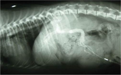
Figure 24.1 Contrast portogram showing extra-hepatic porto-caval shunt entering the caudal vena cava but with partial arborization of the liver.
primary portal vein hypoplasia from portosystemic shunts. Protein C levels were over 70% increased in 88% of cases with primary portal vein hypoplasia and low in dogs with PSS (Gow et al. 2012; Kim et al. 2013).