Diagnosis
Radiographic signs of ILD may be fairly subtle in the face of advanced and severe clinical signs, and often the clinical signs correlate poorly with radiographic severity. In addition, many dogs with ILD are older, and there may be some overlap in lesions from ILDs and age-related changes within the lungs.
Where lesions are readily radiographically demonstrable (Figure 32.1, Figure 32.2); endoscopy and broncho-alveolar lavage (BAL) will likely elucidate the etiology of the lesions. However, it is not uncommon that a patient is fulminant dyspneic with seemingly mild radiographic lesions. In these cases, pulmonary hypertension and pulmonary thromboembolism should be considered. In order to differentiate between these conditions, echocardiography, clotting tests such as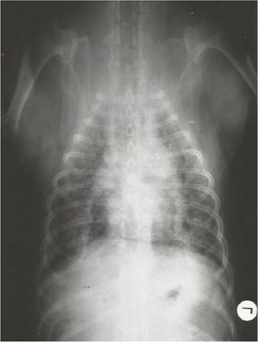
Figure 32.1 Ventro-dorsal thoracic radiograph of 6- year-old dog with pulmonary fibrosis showing advanced interstitial pattern involving all the lung lobes.
thromboelastography and D-dimers, and computed tomography (CT) or CT angiography may be required. In the case of ILDs with subtle radiographic signs, CT may be of great use in diagnosing conditions such as interstitial pulmonary fibrosis (IPF) (Heikkila- Laurila and Rajamaki 2014). Biomarkers such as serum endothelin 1 and procollagen type III aminoterminal propeptide in BAL fluid have demonstrated some utility in differentiating between IPF and chronic bronchitis (Krafft et al. 2011; Heikkila-Laurila and Rajamaki 2014), but to the author's knowledge, these assays are not yet commercially available. A typical diagnostic clinical conundrum is when to recommend a lung biopsy, when an ILD is suspected. Although incontrovertible recommendations cannot be made, a lung biopsy should only be performed once all other reasonable non-invasive options have been exhausted. The author finds it useful to pursue a lung biopsy in cases where immunosuppression is sought as a therapeutic intervention. Given the generally poor response of IPF to immunosuppression, the author would counsel the client against treating IPF aggressively for long periods of time, in the interests of the patient's quality of life, and this discussion is often best guided when informed by a sound histological report.
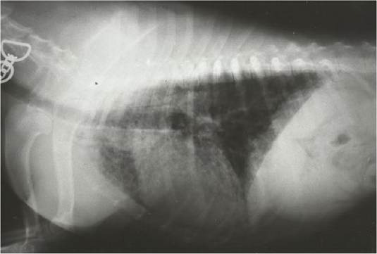
Figure 32.2 Lateral thoracic radiograph of 6-year-old dog with pulmonary fibrosis showing advanced interstitial pattern tending to be bronchial to nodule and involving all the lung fields.