Diagnosis
Laboratory tests are needed to diagnose chytridiomycosis, as tolerant species are infected without being sick, and because in susceptible hosts clinical signs of chytridiomycosis only occur in late terminal stages and are often non-specific.
With heavy infections, changes that are suggestive of Chytridiomycosis may include erythema of ventral surfaces, abnormal posture such as splayed limbs, slow righting reflex and abnormal skin shedding (Fig.
14.2a) (Berger et al. 2009). With Bsal, susceptible salamanders usually develop obvious ulcers (Fig. 14.2b) (Martel et al. 2014). These signs are typical in sick amphibians, and two major diseases that can present similarly are ranaviral disease and bacterial septicemia “redleg.” In tadpoles with Bd, infections cause mouthpart abnormalities including loss of dark tooth rows which can lead to emaciation (Rachowicz and Vredenburg 2004).Details of diagnostic methods for Bd are available in Berger et al. (2009) and the online OIE Manual of Diagnostic Tests for Aquatic Animals 2017 (Chapter 2.1.1), and for Bsal see Martel et al. (2013). A nationwide survey protocol for detecting Bd is also available (Skerratt et al. 2008).
Quantitative polymerase chain reaction (qPCR) is the gold standard for testing. As sampling for PCR involves taking skin swabs, which can be stored at room temperature, this method is non-invasive and is convenient for researchers, veterinarians and conservation managers testing wild or captive animals. For Bd, qPCR was shown to be much more sensitive (72.9%) than histology (26.5%) although was less specific (94.2% versus 99.5%) (Skerratt et al. 2011a, b). Quantitative PCR permits relative quantification of conserved Bd 18S and 28S ribosomal DNA from skin swabs down to a resolution of one genomic zoospore equivalent from as soon as 7 days post infection (Annis et al. 2004; Boyle et al.
2004; Kriger et al. 2006; Hyatt et al. 2007). Standard PCR, however, is also accurate and may be cheaper (Garland et al. 2011). A qPCR to detect Bsal is based on the 5.8S rRNA gene (Martel et al. 2013). Besides, a duplex qPCR detecting Bd and Bsal is available (Blooi et al. 2013, 2016).Microscopy was the original diagnostic method for chytridiomycosis and is highly accurate in sick animals which have heavy burdens. Microscopy includes histology, wet preparations and immunostaining, and requires pieces of whole or shed skin. Also histological examination of all organs by a pathologist is important if ill or dead frogs are found, as part of general disease surveillance (Duffus 2009). For
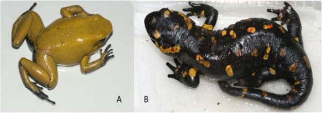
Fig. 14.2 (a) Bd-infected frogs (Phyllobates bicolor) that develop chytridiomycosis show abnormal posture such as splayed limbs and abnormal skin shedding, with remnants of shed skin. (b) After infection with Bsal, susceptible salamanders (Salamandra salamandra) usually develop obvious skin ulcers, here coinciding with haemorrhages (© Pasmans and Martel 2015. All Rights Reserved)
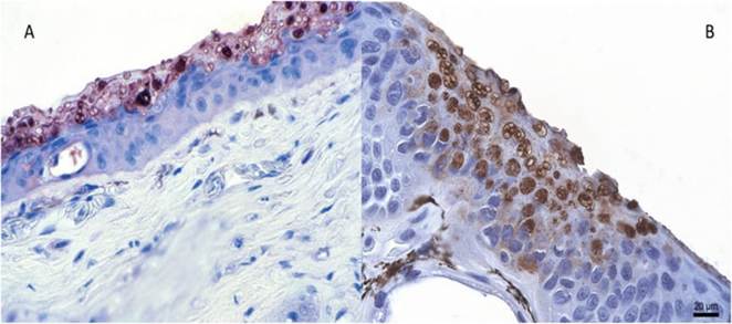
Fig. 14.3 Immunohistochemical staining of the skin of a Bd-infected frog, which died due to Chytridiomycosis (Alytes obstetricans) (reprinted from Berger et al. 2002) (a) and a Bsal-infected fire salamander (b). Bd causes focal epidermal hyperplasia and hyperkeratosis (with abundant thalli present in the keratin), whereas Bsal causes focal necrosis with subsequent erosive and ulcerative lesions. For both chytrids, intracellular thalli (stained dark brown) abound in the lesions (© Pasmans and Martel 2015. All Rights Reserved)
histology, skin from body, digits, or tadpole mouthparts can be examined with haematoxylin and eosin (H&E) staining (Berger et al. 2000, 2009). Sporangia occur in clusters within the epidermis and appear spherical or oval (5-13 μm) with a smooth eosinophilic wall.
Discharge papillae are occasionally seen and project towards the skin surface. Focal hyperkeratosis and erosions are common in Bd- infected areas, whereas Bsal causes necrosis with subsequent erosive and ulcerative lesions (Fig. 14.3). To confirm suspect cases, sporangia can be highlighted using other stains, such as periodic acid-Schiff (PAS) or silver, or an immunoperoxidase stain using polyclonal antibodies with high affinity for Bd and Bsal (Berger et al. 2002; Van Ells et al. 2003; Martel et al. 2013). Wet preparations are quick to prepare using shedding stratum corneum, whole skin or excised tadpole mouthparts that are spread on a slide and cover-slipped (Berger et al. 2009). Diagnosis requires some practice, but the observation of internal septa within sporangia increases confidence in the diagnosis. As Batrachochytrium species are slow-growing compared with microorganism contaminants, culture is difficult and is not used for diagnosis (Berger et al. 2009).14.8
More on the topic Diagnosis:
- Diagnosis
- Diagnosis
- SelectiveIgM Deficiency
- Main Influences on Thyroid Function Tests
- Remission
- Treatment
- Quality ofLife
- Inherited and Congenital Diseases
- Narcolepsy-Cataplexy
- Management