Diagnosis of Hemic-Lymphatic Diseases by Presenting Sign
Bleeding Disorders
Indications of bleeding disorders include petechial or ecchy- motic hemorrhages of mucous membranes, prolonged bleeding from venipuncture sites or surgical wounds, passage of blood from body orifices, or development of subcutaneous or periarticular swellings.
Such signs can result from vasculitis, platelet disorders, or coagulopathies. These categories of disease have received limited attention in goats.Inherited afibrinogenemia has been reported in a family of Saanen goats (Breukink et al. 1972). The inheritance pattern is incomplete autosomal dominant. There is complete absence of circulating fibrinogen in homozygous individuals, and goats thus affected do not live past the kid stage. Unchecked umbilical hemorrhage at birth is the most common presentation, but recurrent hemarthroses and subcutaneous and mucosal hemorrhage can also be seen. Clotting time, thrombin time, stage 1 prothrombin, and partial thromboplastin times are all prolonged in afibrinogenemia. Fibrinogen concentration, as measured by bioassay of thrombin clottable protein, is always under 0.15 g/dL and is usually 0. Acquired coagulation disorders presumably occur in the goat at a rate similar to other species, but the literature on specific causes or cases of coagulopathy in the goat is sparse. Evidence of bleeding from the orifices of a dead goat is suggestive of anthrax.
Thrombocytopenia is reported as a consistent finding in African trypanosomosis in all affected species, including goats (Davis 1982). The degree of thrombocytopenia and the development of subsequent hemorrhage are directly correlated to the degree of parasitemia that develops.
Bracken fern (Pteridium aquilinum) ingestion by cattle can lead to a syndrome of pancytopenia with leukopenia, thrombocytopenia, and anemia. Melena, epistaxis, and widespread petechial and ecchymotic hemorrhage are major clinical findings.
While the morbidity rate may be low, the mortality rate is high. There is one report of naturally occurring bracken fern toxicity in goats, but no signs of hemorrhage were observed (Tomlinson 1983). Bracken fern has been linked epidemiologically to esophageal and stomach cancers in humans and the plant is known to contain a carcinogen, ptaquiloside. Ptaquiloside has been detected in the milk of cows fed on a diet containing bracken fern and also has been identified in pooled samples of raw goat milk from flocks grazing on pastures containing bracken fern (Virgilio et al. 2015).Hemorrhagic diathesis is considered to be a clinical manifestation of severe, diffuse liver disease in other farm animal species (Constable et al. 2017). However, a review of liver diseases of sheep and goats did not identify coagulopathy as a clinical outcome of hepatic disease (Fetcher 1983). The only report documenting decreased clotting activity in goats in association with liver disease involved experimental dosing with carbon tetrachloride (Jones and Shah 1982).
Anemia
Anemia is suggested clinically by pale or white mucous membranes (Figure 7.1), exercise intolerance, tachypnea, tachycardia, possible systolic murmurs, weakness, and (in extreme cases) collapse. When anemia is a result of
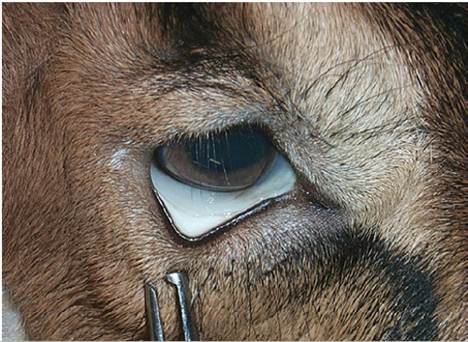
Figure 7.1 White-colored conjunctiva characteristic of a goat with marked anemia. In normal goats, the color is pink to red. Source: Courtesy of Dr. M.C. Smith.
intravascular hemolysis, then jaundice and hemoglobinuria are also important clinical signs. Signs of anemia are frequently accompanied by signs of hypoproteinemia, particularly intermandibular edema, ascites, and weight loss. Anemia is a common and important clinical presentation in goats.
Causes of Hemolytic Anemia
Important, established causes of hemolytic anemia in goats include the hemoparasitic diseases anaplasmosis, babesiosis, eperythrozoonosis, and theileriosis; nutritional disorders including copper toxicosis, kale ingestion, and consumption of other, regional poisonous plants; and an infectious cause, leptospirosis.
Other suspected causes of hemolytic anemia in goats include infections due to Clostridium novyi type D (Cl. hemolyticum) and Clostridium perfringens type A, as reported in sheep. Experimental poisoning with oak tannins caused marked hemolytic anemia in goats, but naturally occurring oak poisoning is uncommon in this species (Begovic et al. 1978). Experimental infections of sarco- cystosis (sarcosporidiosis) produce hemolytic anemia (Dubey et al. 1981), but almost all known naturally occurring infections in goats are subclinical, producing muscle cysts seen at slaughter or necropsy, as discussed further in Chapter 4.
Two reports suggest hypophosphatemia as the cause of hemolytic anemia and hemoglobinuria in female goats, but in two of the three reported cases, serum inorganic phosphorus levels were in the normal range (Setty and Narayana 1975; Samad and Ali 1984). In cattle with postparturient hemoglobinuria, serum inorganic phosphorus levels are well below the normal range.
Causes of Blood Loss Anemia
Anemia due to loss of blood is of major clinical significance in goats. The condition is most often associated with some form of parasitism. Important causes of blood loss anemia include infestations by Haemonchus spp. and liver flukes, especially Fasciola hepatica. External parasitic causes include sucking lice, ticks, and fleas (Schillhorn van Veen and Mohammed 1975). Predation may be another important cause of blood loss. While wild predators can be expected to kill goats outright unless interrupted, domesticated dogs often maim goats without killing them. Severe hemorrhagic trauma often results.
Causes of Anemia Due to Impaired Erythropoiesis
Anemias of this type occur infrequently or are overshadowed by other concurrent and more prominent clinical signs. Nutritional causes include cobalt, copper, and iron deficiencies. Toxic causes include fluorosis and possibly bracken fern ingestion. Anemia of chronic infection also occurs in goats (e.g., in paratuberculosis).
Iron deficiency is associated with prolonged feeding of doe's milk to kids without mineral supplementation or access to forage. Copper deficiency manifests primarily as a neurologic disease in young kids. In experimentally induced cobalt deficiency in goats, a macrocytic, normochromic anemia was observed in addition to weight loss (Mgongo et al. 1981). In naturally occurring cases of cobalt deficiency, ill-thrift is a consistent finding, but the presence of anemia is variable (Brain 1983; Black et al. 1988). Non-regenerative anemia has been documented in chronic fluorosis of goats grazing near a superphosphate factory in Egypt (Karram et al. 1984). One incident of bracken fern poisoning has been reported in goats. Anemia was present, but may have been due to concurrent parasitism (Tomlinson 1983).
There is one report of myelofibrosis occurring in 11 pygmy goat kids from two litters with a breeding history that suggested that the condition was inherited. Affected kids were lethargic, had poor appetite, pale mucous membranes, and died by 12 weeks of age. Hematologic examination revealed severe anemia, moderate leukopenia, severe neutropenia, and reticulocytosis. At necropsy, marked myelofibrosis was noted, especially in diaphyseal bone marrow samples, and there was evidence of active extramedullary hematopoiesis at multiple sites, including thymus gland, tonsils, lymph nodes, liver, and elsewhere (Cain et al. 1994).
Documented causes of anemia in goats are summarized in Table 7.6. The table emphasizes concurrent clinical and laboratory findings, such as hypoproteinemia and hemoglobinuria, which can aid in the differential diagnosis of anemia. All the hemoparasitic diseases, leptospirosis, copper poisoning, phosphorus deficiency, kale ingestion, and other poisonous plants causing anemia, are discussed in detail later in this chapter. Other diseases associated with anemia are discussed elsewhere in the text because other clinical signs predominate.
Lymphadenopathy
Transient swelling of select regional lymph nodes can be expected in common, localized infections such as mastitis, or subsequent to vaccinations.
Persistent lymphadenopathy, however, is a major clinical finding in many important caprine diseases, including caseous lymphadenitis, theile- riosis, trypanosomosis, melioidosis, tuberculosis, nocardiosis, and lymphosarcoma. Theileriosis and trypanosomosis are discussed in detail in this chapter because of the significant role of anemia in these diseases. The remainder of the diseases are discussed in Chapter 3.Table 7.6 Anemia in goats: aids for differential diagnosis.
| Cause of anemia | Pathogenesis and morphologic type | Role of anemia in disease | Total serum protein | Icterus (jaundice) | Hemoglobinuria | Other clinical signs | Comments |
| Anaplasmosis | Hemoparasitic; extravascular hemolysis; regenerative | Major | Normal | Likely | No | Few; abortions can occur, concurrent disease common | Often subclinical |
| Babesiosis | Hemoparasitic; intravascular hemolysis | Major | Normal | Likely | Likely | Fever, diarrhea, abortion | Poorly described in goats |
| Theileriosis | Hemoparasitic; pathogenesis of anemia unclear | Minor | Normal | Variable | Transient | Fever, swollen lymph nodes, lacrimation | Primarily a parasite of white cells |
| Trypanosomosis | Hemoparasitic; mainly extravascular hemolysis | Major | Normal to low | Unlikely | Unlikely | Fever, edema, lymphadenopathy, weight loss | Seen primarily in Africa |
| Eperythrozoonosis | Hemoparasitic; mainly extravascular hemolysis | Major | Normal | Likely | No | None, but concurrent disease common | Poorly described in goats |
| Leptospirosis | Septicemia; intravascular hemolysis | Major | Normal | Yes | Yes | Fever, abortion | Uncommon in goats |
| Copper poisoning | Nutritional; intravascular hemolysis | Major | Normal | Yes | Yes | Acute death | Goats more resistant than sheep |
| Haemonchus spp. | Gastric parasite; blood loss anemia | Major | Low | No | No | Weight loss, edema | Major cause of anemia in goats |
| Coccidiosis | Intestinal parasite; blood loss anemia | Major | Low | No | No | Diarrhea or dysentery; dehydration | Especially young goats affected |
| Liver flukes | Liver parasite; blood loss anemia | Major | Low | Likely | No | Weight loss, edema, ascites, eosinophilia | |
| Schistosomosis | Vascular parasite; blood loss anemia | Major | Low | No | No | Weight loss, diarrhea, ascites | Numerous spp. affect the goat |
| External parasites (ticks, fleas, lice) | Skin parasites; blood loss anemia | Usually minor | Normal to low | No | No | Pruritus, rough hair coat | Sucking lice and fleas may cause severe anemia |
| Trauma/predation | Blood loss anemia | Minor | Normal to low | No | No | Shock, musculoskeletal | Predation a serious problem |
| Cobalt deficiency | Nutritional; red blood cell multiplication reduced; macrocytic anemia | Minor | Normal to low | No | No | Weight loss, diarrhea, lacrimation, weakness | Mimics gastrointestinal parasitism |
| Cause of anemia | Pathogenesis and morphologic type | Role of anemia in disease | Total serum protein | Icterus (jaundice) | Hemoglobinuria | Other clinical signs | Comments |
| Copper deficiency | Nutritional; reduced heme synthesis; microcytic anemia | Minor | Normal | No | No | Enzootic ataxia, or “swayback” | Primarily neurologic; mostly in young goats |
| Iron deficiency | Nutritional; reduced heme synthesis; microcytic anemia | Minor | Normal | No | No | None | Uncommon; seen in milk-fed kids |
| Phosphorus deficiency | Nutritional; hemolytic anemia | Major | Normal | Yes | Yes | None | Some cases reported from India |
| Chronic diseases (e.g., paratuberculosis) | Anemia of chronic disease, non- regenerative | Minor | High to low | No | No | Weight loss, possible edema | |
| Kale poisoning | Plant toxicity; Heinz body anemia; regenerative | bgcolor=white>Major Normal | Likely | No | None | Goats more resistant than cattle | |
| Myelofibrosis | Inherited (?) | Major | Normal | No | No | Lethargy, inappetance | Rare |