Diagnostics
As with any disease process, diagnosis of a GB MC requires assimilating data from the signalment of the patient, owner history, clinical signs, clinical examination findings, laboratory data, and diagnostic imaging findings.
GB MCs commonly present in middleaged to old dogs, with no definitive breed predisposition although they are overrepresented in Shetland Sheepdogs, Cocker Spaniels, Miniature Schnauzers and Dachshunds. There is no apparent sex predilection. Because MCs are thought to develop slowly; presenting clinical signs are variable with a waxing and waning nature. In fact, MCs are often detected as incidental findings on routine abdominal ultrasonography when the animal is showing no clinical signs of systemic disease. In the majority of cases subacute to chronic non-specific, vague clinical signs are seen including vomiting, anorexia, lethargy, diarrhea, polyuria-polydipsia, icterus, dehydration, tachycardia, tachypnea, and abdominal discomfort. Alternatively, patients may present with signs of acute abdomen associated with extrahepatic bile duct obstruction, pancreatitis or bile peritonitis. Pyrexia, if present, is often associated with bacterial cholecystitis or bile peritonitis.
Routine clinico-pathological abnormalities seen are indistinguishable from other hepatobiliary diseases and certain endocrinopa- thies and are not particularly useful in making a diagnosis. These include increases in serum liver enzyme activity (ALP, ALT, and GGT), hyperbilirubinemia, hypertriglyceridemia, and hypercholesterolemia. Neutrophilic leucocytosis, often with a left shift, can occur with bile peritonitis and bacterial cholecystitis.
Abdominal radiographs are not adequate for making a definitive diagnosis but are useful for excluding other causes of acute abdomen or vomiting such as gastrointestinal obstruction, or abdominal masses and other hepatobiliary diseases including acute liver injury, neoplasia, and choleliths.
Abdominal ultrasound is the preferred and most sensitive diagnostic test for GB MCs, which have distinct ultrasonographic characteristics. The tenacious mucin along the GB wall is visualized as a hypoechoic rim. The central contents are hyperechoic and non-gravity dependent with ballotte- ment or re-positioning of the patient. Radiating striations extend from the GB wall centrally as the MC matures, creating the described “kiwi-fruit” appearance (Figure 22.1) (Besso et al. 2000, Hottinger 2014). Ultrasonography may also detect signs of extrahepatic biliary obstruction as the mucinous bile extends into the cystic, common, and hepatic ducts. Ultrasonographic features of GB wall necrosis
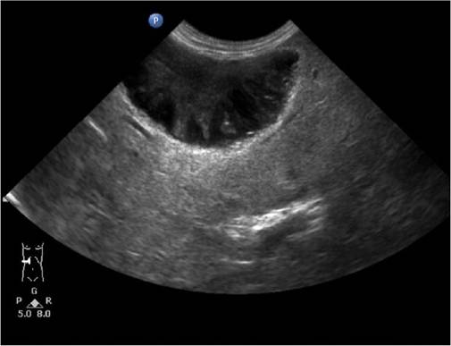
Figure 22.1 Longitudinal ultrasound image of a gallbladder mucocele demonstrating the characteristic "kiwi-fruit" or stellate appearance. The mucin layer is hypoechoic while the hyperechoic bile is centrally located. (Courtesy Dr R. Friedlein, Fourways Veterinary Hospital, South Africa.)
or early rupture of the GB include an irregular outline or defect to the GB wall, hyperechoic surrounding mesenteric fat or a hypoechoic halo of fluid surrounding the GB itself (Besso et al. 2000). One disadvantage of ultrasonography in these cases however, is that it lacks the sensitivity to accurately assess the presence of pressure necrosis of the GB wall or cystic duct which can lead to eventual rupture and subsequent bile peritonitis in some cases.
Because historical and presenting signs are often so vague and laboratory abnormalities seen so non-specific, often many animals are presumed to be suffering from other medical conditions such as pancreatitis or chronic gastrointestinal diseases such as IBD, especially when diagnostic imaging tools such as abdominal ultrasonography are not readily available to the clinician. It is, therefore, imperative that the clinician carefully reviews all available information and refers cases to institutes where ultrasonography is available to achieve an accurate and timely diagnosis. Often however, a diagnostic conundrum can occur when other concomitant disease processes such as pancreatitis or cholangio- hepatitis are present. In these cases, it becomes a diagnostic challenge to decide whether the GB MC present is an incidental finding or contributing to the clinical signs seen.