Esophagitis
Esophagitis is an acute or chronic inflammatory disorder of the esophageal mucosa. If severe, esophageal inflammation may extend into the submucosal and muscular layers. Esophagitis can result from ingestion of corrosive or caustic substances, foreign body obstruction, thermal burns, infectious agents, persistent vomiting, and probably most commonly, gastroesophageal reflux (GER).9-14 The esophageal mucosa has a barrier function to prevent damage by various substances, including gastric acid.
This barrier function is provided by the stratified squamous epithelium, tight junctions between the epithelial cells, the mucus layer, prostaglandin production, surface bicarbonate ions, and peristaltic clearance of material from the esophagus.15The clinical signs are often related to the severity and depth of inflammation. Characteristic signs include dysphagia, ptyalism, regurgitation, gagging, varying degrees of inappetence, repeated swallowing motions with extension of the head and neck, odynophagia, lethargy, and weight loss.11,13,14 The physical examination is often unremarkable, but may show evidence of aspiration pneumonia (i.e., fever and/or increased lung sounds). Diagnostics generally include a minimum database (CBC, serum chemistry profile, urinalysis, and thoracic and/ or abdominal radiographs), which is generally unremarkable. The CBC may show a leukocytosis if severe esophagitis or aspiration pneumonia is present. A barium esophagram performed under fluoroscopy may show GER, segmental dilation of the esophagus, an irregular esophageal mucosa, and /or decreased esophageal motility.13,14 A definitive diagnosis is made via endoscopy and histopathology. At endoscopy, the mucosa may appear hyperemic and irregular, and polypoid masses, nodules, and/or ulcerations with spontaneous bleeding may be seen (Figure 3.3).11,13,14,16 Loss of distensibility of the esophagus is characteristic for chronic esophagitis.16 The esophagus can be difficult to biopsy with standard instruments due to its tough stratified squamous epithelium.
However, abnormal, eroded, ulcerated, or proliferative esophageal lesions can generally be biopsied easily.14,16 Histopathology may show an eroded, ulcerated, hyperplastic, or dysplastic epithelium, and /or lymphocytic, plasmacytic, or neutrophilic infiltrates of the submucosa.13,14Mild esophagitis may be treated conservatively by withholding food for 2-3 days. Moderate to severe esophagitis should be treated more aggressively. Withholding food fed by mouth
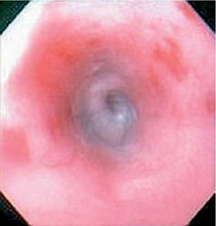
Figure 3.3:
Esophagitis. Endoscopic view of the distal esophagus of a 5-year-old spayed female DSH with protracted vomiting of 5 days' duration. Note the areas of hyperemia and erosion indicating esophagitis.
and providing nutrition via a gastrostomy tube has been recommended for patients with moderate to severe acute esophagitis in the past.17 However, some authors recommend continuing to feed the animal by mouth throughout treat- ment.13,14,18 Whether to place a gastrostomy tube should be decided on an individual basis. Those animals that are completely anorectic, in poor body condition, and those with esophagitis that is in danger of perforating should receive a gastrostomy tube.
Various medical therapies are also recommended. Oral sucralfate suspension has been noted to be one of the most important therapies.17 Sucralfate has cytoprotective properties when bound to an eroded or ulcerated site.19,20 However, there are no clinical studies which show that sucralfate is effective in patients with esophagitis. Gastric acid suppression is also recommended. This may be accomplished either using an H2- receptor antagonist (e.g., cimetidine, ranitidine, or famotidine) or a proton pump inhibitor (e.g., omeprazole). The choice generally depends on the severity of the esophagitis and whether continued GER is present. Proton pump inhibitors may be advantageous in patients with severe erosive esopha- gitis.21 Prokinetic agents are also recommended in order to increase lower esophageal sphincter tone and promote gastric emptying, thus decreasing the amount of GER.12-14 Metoclopramide and cisapride have been used for this purpose. Since cisapride has been removed from the market due to side-effects in humans, it may be difficult to obtain for veterinary patients. However, several compounding pharmacies offer cisapride. Ranitidine and nizatidine have been shown to stimulate gastrointestinal motility by inhibiting acetylcholinesterase activity and may be useful for promotion of gastric emptying in patients with esophagitis.22
3.3.3