Etiology and Taxonomy
The genus Cryptococcus (teleomorph Filobasidiellai) comprises basidiomycetous yeast species, most of which are environmental saprophytes that do not cause infections in human or animals (Kwon-Chung et al.
2017). The pathogenic agents of cryptococcosis are classified into two species, C. neoformans and C. gattii (Table 12.1). The species C. neoformans comprises two varieties, C. neoformans var. grubii and C. neoformans var. neoformans (Meyer et al. 2009). The species C. neoformans consists of the VNI-VNIV and VNB molecular genotypes, comprising var. grubii (serotype A or VNI, VNII, and VNB strains), var. neoformans (serotype D or VNIV strains), and serotype AD strains (VNIII), which represents hybrids of the two varieties (Kwon-Chung and Varma 2006). The species C. gattii is subdivided into two serotypes (B and C) and four molecular types (VGI, VGII, VGIII, and VGIV) (Boekhout et al. 2001). Diseases caused by other Cryptococcus species, such as Cryptococcus laurentii and Cryptococcus albidus, have been reported infrequently and generally in immunocompromised hosts (Harris et al. 2012).Recently, by combining various molecular genotyping methods, a new taxonomical classification system, based on a seven species/four hybrid scheme, has been proposed for pathogenic cryptococcal species (Hagen et al. 2015). This scheme includes the following species: C. neoformans [C. neoformans Cryptococcus neoformans var. grubii (serotype A) with three genotypes (VNI, VNII, and
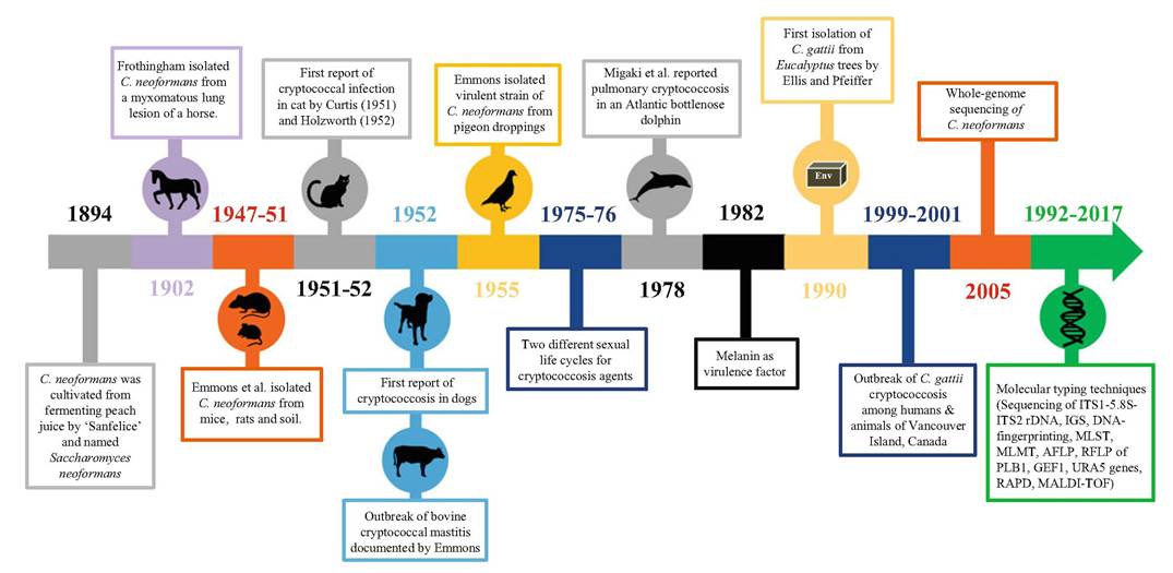
Fig. 12.1 The main facts and pioneering events that contributed to the epidemiological studies on human and animal cryptococcosis. Env environment, MLEE multilocus enzyme electrophoresis, RAPD random amplification of polymorphic DNA, AFLP PCR-Angerprinting amplified fragment length polymorphism, RFLP restriction fragment length polymorphism, rDNA ribosomal DNA region, IGS intergenic spacer region, MLST multilocus sequence typing, MLMT multilocus microsatellite typing, MALDI-TOF matrix-assisted laser desorption ionization-time-of-flight mass spectrometry-based method analysis
252 K.
Singhetal.Table 12.1 Classification of Cryptococcus neoformans and Cryptococcus gattii species complexes
| Species complexes | Molecular type | |
| Cryptococcus neoformans | Cryptococcus neoformans var. grubii | vnwniwnb (aflpi,ia/ 1B, VNB) |
| Cryptococcus neoformans var. neoformans | VNIV (AFLP2) | |
| Cryptococcus neoformans AD hybrid | VNIII (AFLP3) | |
| Cryptococcus gattii | Cryptococcus gattii | VGI (AFLP4) |
| VGII (AFLP6) | ||
| VGIII (AFLP5) | ||
| VGIV (AFLP7) | ||
| VGIV/VGIIIc (AFLP10) | ||
| Cryptococcus gattii DB hybrid | AFLP 8 | |
| Cryptococcus gattii AB hybrid | AFLP 9 | |
| Cryptococcus gattii AB hybrid | AFLP 11 | |
VNB)], C. deneoformans [C. neoformans var. neoformans (serotype D or VNIV)], 5 C. gattii cryptic species with serotypes B/C or VGI-IV [C. gattii (VGI), C. bacillisporus (VGIII), C. deuterogattii (VGII), C. tetragattii (VGIV), and C. decagattii (VGIV/VGIIIc)], as well as the following hybrids, C. neoformans x C. deneoformans hybrid, C. deneoformans x C. gattii hybrid, C. neoformans x C. gattii hybrid, and C. deneoformans x C. deuterogattii hybrid. However, a panel of experts representing numerous laboratories around the world agreed that in the absence of biological differences among clades, a new nomenclature system for Cryptococcus was preferable to naming each clade as a separate species. Accordingly, “C. neoformans species complex” and “C. gattii species complex” were both proposed (Table 12.1), as the etiological agents of cryptococcosis (Kwon-Chung et al. 2017). Furthermore, a new hypothesis also suggested continental drift as a possible speciation driver within the Cryptococcus species complexes, adding greatly to the ongoing discussion of speciation between these complexes (Casadevall et al. 2017). On the basis of the geographic distribution of these two species complexes and the coincidence of the evolutionary divergence and Pangea breakup times, it was proposed that a spatial separation caused by continental drift resulted in the emergence of the C. gattii and C. neoformans species complexes from a Pangean ancestor (Casadevall et al. 2017).
12.3