Introduction
Dermatophytes are the most successful pathogenic fungi causing superficial mycoses (dermatophytosis, also called ringworm) in humans and animals. They encompass ecologically and phylogenetically related fungi belonging to the family Arthrodermataceae (order Onygenales) which are able to use keratin as a sole nutrient source (Graser et al.
1999a; Sugiyama et al. 2002). In the last several decades, the dermatophytes were usually categorized into three genera, Trichophyton, Epidermophyton, and Microsporum, and associated sexual state used to be classified in Arthroderma (Weitzman et al. 1986; Weitzman and Summerbell 1995). With complete abolition of dual nomenclature (McNeill et al. 2012) and availability of multiple gene phylogenies, the number of genera was expanded (de Hoog et al. 2017), but the most important primary pathogenic species remains in the three mentioned genera. Chronology of selected important historical events related to dermatophytosis and dermatophytes is shown in Fig. 3.1.Dermatophytoses occur frequently in livestock, in companion animals, and also in wildlife. Infections caused by zoophilic dermatophytes are usually benign and self-limiting and respond well to treatment; they are rarely serious or manifest as systemic infection in immunocompromised host (Chermette et al. 2008; Rouzaud et al. 2016). The prevalence in animals shows large geographic differences. It seems to be influenced by trade, exchanges of animals for reproductive purposes, exhibitions (for cats and dogs), and sportive activities (e.g., horse races). Risk of zoonotic transmission depends on the local spectrum of kept animals and epidemiological situation (prevalence of pathogens in animals) but also on local relations between man and animals, hygienic standards, and socioeconomic factors. Dermatophytoses are highly contagious, and animals kept in herds or groups are threatened by the epidemic spread of infection even when one of few infected individuals are introduced into the community. The environment contaminated by arthroconidia and diseased animals (commonly with mild clinical signs or symptomless) represents a potential source of infection to humans.
High financial costs are associated worldwide with treatment, diagnosis, and prevention of dermatophytosis (Drake et al. 1996). Direct economic costs in farming and industry result from unaesthetic aspect of lesions (hide and skin industry), which
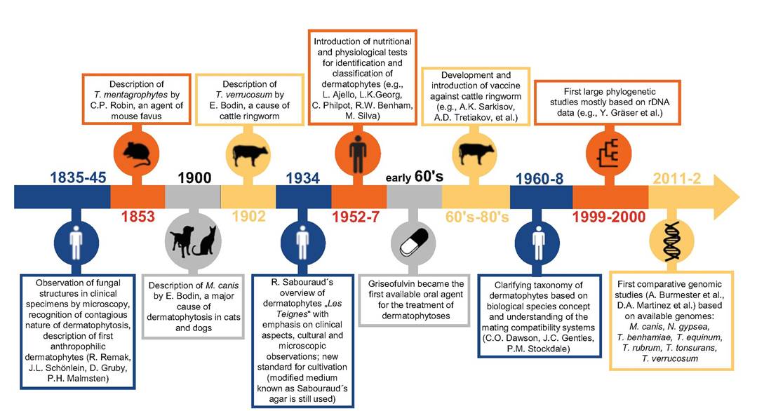
Fig. 3.1 Chronology of important events related to dermatophytosis and taxonomy of dermatophytes
hinder animal trade and are also an obstacle to attend exhibitions and sportive activities (Chermette et al. 2008).
For effective treatment and prevention of the spread of these diseases, it is important to correctly determine the causal agents at the species level, which allows prescription of suitable therapy and at the same time identification of probable source of infection. Correct species identification is an indispensable prerequisite for monitoring changes in the frequency of individual species, helps to evaluate the results of preventive measures and interventions, and is a basic requirement for the preparation of epidemiological studies. Instable taxonomy of dermatophytes, problematic species concept, and phenotypic identification limit comparability of recent epidemiological data with studies from the past. In the last two decades, the advent of molecular methods greatly contributed toward the understanding of the biodiversity of dermatophytes (Graser et al. 1999b, 2000a, 2000b, 2008; Hubka et al. 2014a), thus challenging the previous classification based on morphology and nutritional tests. However, dermatophytosis in animals is classically based on phenotypic methods, and we have currently no systematic data supported by molecular methods worldwide on the epidemiology of dermatophytes.
3.2