Introduction
By far the most important diagnostic tool that veterinarians can utilize is their ability to obtain a complete history and conduct a thorough physical examination. The objective of the physical examination is to recognize and describe gross deviations of the patient’s physical appearance and behavior from those recognized as normal for the animal’s species, breed, age, sex, and sexual status.
The trend toward an increased use of laboratory tests and instrumentation has added diagnostic capabilities, but such techniques are useful adjuncts for diagnosis only when a careful physical examination has been carried out. Thus, information gathered from laboratory and diagnostic imaging procedures must be considered a supplement to, but not a substitute for, hands-on examination. Inspection of the patient, palpation, percussion, and auscultation, all have a place in every examination. Only acute life-threatening situations require a shorter initial examination, until the animal’s condition can be stabilized. In fact, if the patient is in shock or hemorrhaging, or has gastric dilation/volvulus, it is essential to initiate supportive care immediately and obtain a complete history with careful examination afterwards.
The physical examination of the gastrointestinal tract, as described in this chapter, is part of the methodical and standardized physical examination first developed at the School of Veterinary Medicine of Bologna University.1
In this chapter, the discussion is mainly limited to parameters of the physical examination that are specific and directly related to the gastrointestinal system, but it is important to note that the clinician should examine all body systems when presented with an patient that manifests signs of gastrointestinal disease. Ophthalmic and neurological examination, not included in this chapter, should not be forgotten because they can sometimes provide invaluable clues to the cause of a gastrointestinal dysfunction.
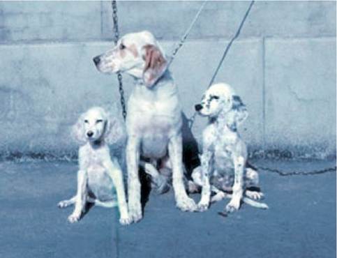
Figure 1.6:
Exocrine pancreatic insufficiency. This picture shows the failure to grow in two puppies with exocrine pancreatic insufficiency compared with a healthy littermate shown in the middle (these dogs are described in detail in: Boari A. et al. Observations on exocrine pancreatic insufficiency in a family of English setter dogs. J Small Animal Practice 1994, 35: 247-250).
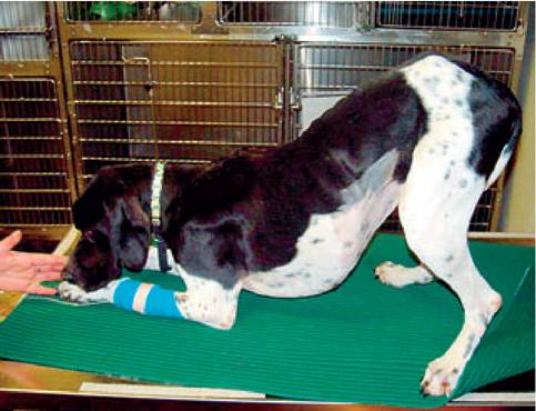
Figure 1.7:
“Prayer” position. This picture shows a dog with acute abdominal pain due to acute pancreatitis. This dog has assumed the “prayer” position with his front legs and sternum on the floor and his hind legs standing.
1.2.2