Intubation
Endotracheal intubation with a cuffed tube is advisable whenever general anesthesia is induced in an adult goat, to prevent inhalation of saliva and regurgitated rumen contents (Taylor 1991).
A variety of rapidly acting drugs may be used to facilitate intubation. These include short-acting barbiturates, ketamine, xylazine, and propofol parenterally and halothane, isoflurane, or sevoflurane via mask. IM administration of midazolam at 0.4 mg/kg followed by ketamine at 4 mg/kg IV is adequate for intubation (Stegmann 1998). Another option is propofol (Waterman 1988) at 3-4 mg/kg slowly IV (Taylor 1991). Ketamine and propofol can be combined for induction in order to reduce the risk of apnea.Laryngospasm has been reported, especially during intubation of young goats. This potentially fatal complication can be prevented by spraying the larynx with a local anesthetic at least 30 seconds before attempting intubation (Taylor 1991). Lidocaine is preferred to benzocaine, to avoid methemoglobinemia (Riebold 2015). The endotracheal tube should be left in place until coughing and swallowing reflexes are regained.
Endotracheal Tube Size
The recommendations given in Table 17.2 aid in the selection of an appropriate tube (Linzell 1964; Gray and McDonell 1986b). A 35 cm long tube is appropriate for an
| Table 17.2 Endotracheal tube sizes for goats. | |
| Weight of goat | Tube size |
| 15 kg | 5-6 mm |
| 25 kg | 7-8 mm |
| 30-40 kg | 9-10 mm |
| Adult dairy goat | 11-12 mm |
Source: Based on Linzell 1964; Gray and McDonell 1986b.
adult goat of a European breed. For all breeds, tubes can be sized externally with reference to the carina of the trachea. Care should be taken to choose a tube of appropriate length and not insert past this premeasured level. Cuffs should be inflated for at least five minutes before induction to detect any slow leaks. It is helpful to stiffen the tube with a wire stylet, which is withdrawn when the tube is in the trachea. Alternatively, a human endotracheal tube exchanger can be preplaced and the endotracheal tube advanced over it (Bush 1996). Both lung fields can be ausculted after tube placement to verify endotracheal versus endobronchial intubation. After intubation, a wooden bite bar can be taped in place to protect the tube.
Intubation under Visual Observation
Direct observation with a laryngoscope is considered ideal for intubation. The anesthetized goat is left in sternal recumbency, and the head is directed toward the ceiling or the goat is placed on its back with its head and neck fully extended. The latter procedure theoretically increases the risk of regurgitation occurring before intubation can be completed. An assistant holds the tongue out of the mouth using a gauze sponge for a better grip. Gauze loops also allow an assistant to hold the jaws wide open without being in the way (Figure 17.1). The lower gauze loop could go over the tongue. The anesthetist passes a laryngoscope with a long blade (20-27.5 cm) to behind the base of the tongue, then lifts the blade to expose the larynx and vocal cords. If a long laryngoscope blade is not available, one can be prepared by welding an extension onto a shorter blade to achieve a total length of 25.7 cm or longer (Tillman and Brooks 1983), although an alternative light source may be necessary. It is also possible to pass a lubricated
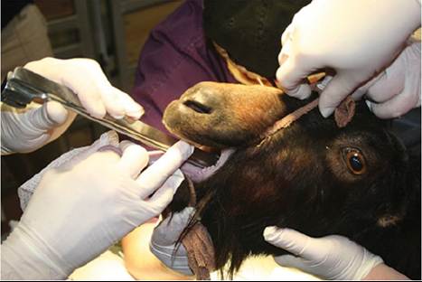
Figure 17.1 Visualizing the larynx with a long laryngoscope blade.
Gauze loops hold the goat's mouth open. Source: Courtesy of Dr. M.C. Smith.endotracheal tube via the inferior nasal meatus, although the tube diameter must be smaller than is listed in Table 17.2 and intubation forceps may be needed (Clarke et al. 2014).
Blind Intubation
Blind intubation is worth learning, not only because laryngoscopes are expensive, but also because the batteries lose their charge. The goat is placed in lateral recumbency with the head in slight extension, in line with the thoracic spine. The thumb and first finger of one hand hold the tongue and lower jaw, while the other fingers of the hand press against the hard palate to hold the mouth open. The endotracheal tube is held with the other hand and passed into the pharynx with its concave side against the tongue until its tip is felt to touch the larynx. The tube is then twisted and advanced until the characteristic feel of its passage over the tracheal rings is detected (Hecker 1974).
Others prefer to advance the tube into the pharynx with its convex surface against the tongue, then to rotate the tube 180° to ensure that the tip of the tube is not trapped beneath the epiglottis. With this technique, which has been well illustrated by radiographic studies, the larynx is pushed upward with one hand to occlude the esophagus while the other advances the tube (Gray and McDonell 1986b).