Late Abortion: Infectious Causes
In the United States, chlamydiosis, Q fever, and toxoplasmosis appear to be the most frequently identified infectious causes of caprine abortion. Other agents may cause serious losses in individual herds and vaccination programs may affect the relative importance of some diseases.
Listeriosis was the most common cause of abortions in goats in the Netherlands in the 2012-2013 kidding season (van Engelen et al. 2014); that paper reviews reports from other countries as well. Additional literature has been reviewed by Lefevre (1987). The zoonotic potential of most of these agents should not be forgotten.Akabane, Cache Valley, and Schmallenberg Viruses
Akabane virus has caused abortions and perinatal mortality of calves, lambs, and kids (Markusfeld and Mayer 1971; Inaba et al. 1975; Kirkland 2015).
Etiology and Epidemiology
Akabane virus is an arbovirus of the bunyavirus group. The virus is arthropod borne, by gnats and mosquitoes. The disease has been recognized in Africa, the Middle East, Southeast Asia, and Australia (Kirkland 2015). It is exotic to the United States. Outbreaks are believed to occur when susceptible females are in early pregnancy during periods of increased vector activity. As many as 50% of kids born on some farms have been affected (Shimshony 1980).
Pathogenesis
The pregnant dam develops viremia. Akabane virus infects lambs transplacentally at 30-36 days of gestation (Parsonson et al. 1975, 1981). The critical time for infection of kids is probably similar (Kurogi et al. 1977). At this early period, the virus destroys embryonic cells in the cerebral cortex. Fetal muscles undergo neurogenic atrophy, as discussed in Chapter 4. Fetal death and mummification may occur. If the fetus survives, the virus is cleared, but precolostral antibodies remain as evidence of the infection.
Clinical Signs
The disease is sometimes referred to as congenital arthro- gryposis-hydranencephaly syndrome.
Malformations associated with Akabane virus include micrencephaly, hydrocephalus, hydranencephaly, porencephaly, arthrogryposis, and reduced muscle mass (Haughey et al. 1988). Fetuses may be stillborn or die very soon after birth. Arthrogryposis or prepartum death of the fetus may contribute to dystocia. The pregnant dam shows no overt clinical signs at the time of initial infection.Similarity to Cache Valley Virus,
Schmallenberg Virus
A similar arthropod-borne bunyavirus, the Cache Valley virus, is endemic to parts of North America and has caused epizootics of arthrogryposis with hydrocephalus, hydra- nencephaly, or cerebellar dysplasia in lambs in Texas, Michigan, and Nebraska (Edwards et al. 1989; Chung et al. 1990b; de la Concha-Bermejillo 2003) and elsewhere. A high prevalence of mummified fetuses, stillbirths, and weak lambs has been observed in infected flocks and reproduced experimentally by inoculation at 27-54 days of gestation (Chung et al. 1990a). Oligohydroamnion contributes to lack of fetal movement and consequent arthrogryposis.
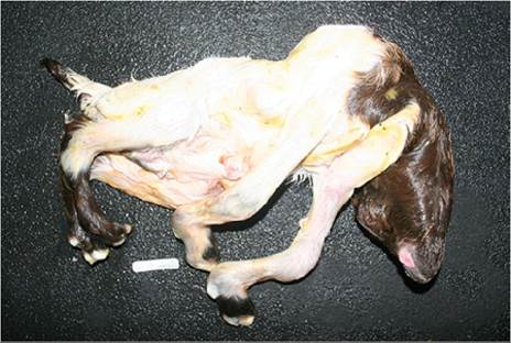
Figure 13.5 Ventral view of an arthrogrypotic Boer kid documented to have been infected with Cache Valley virus by the presence of antibodies in pleural fluid. Source: Courtesy of Dr. M.C. Smith.
Although case reports are rare, goat fetuses are also susceptible to Cache Valley virus (Edwards et al. 2003; Harvey et al. 2019). An outbreak in New York in December of 2015 resulted in three abortions and the delivery of deformed kids from four other does (see Figure 13.5). Some of these kids had shortened hindlimbs while the forelimbs were in rigid extension, along the body, resulting in dystocia.
Identical malformations were first seen in Europe in 2011 in calves, lambs, and goat kids, caused by another orthobunyavirus, the Schmallenberg virus (Herder et al. 2012; Beer et al. 2013). Relatively few goats were affected, possibly because they were less likely to be housed outdoors where they might be exposed to the Culicoides vector during early pregnancy (Helmer et al.
2013). Similarly, the Shuni virus in Israel (Golender et al. 2015) has caused malformations in goat kids.Diagnosis and Control
Congenital malformations, as described above, are suggestive of bunyavirus infections. Retrospectively, the first trimester of pregnancy will have occurred during a period of biting insect activity, especially Culicoides gnats and mosquitoes. A warm, humid period after several years of drought results in a large population of insect vectors interacting with a naive population of small ruminants. Adjusting the breeding period to avoid times of high vector activity may be helpful.
Virus isolation may be attempted from placenta, fetal muscle, or fetal brain, but is unsuccessful in full-term fetuses. Polymerase chain reaction (PCR) tests have also been developed for these viruses. Demonstration of preco- lostral or fetal serum antibodies against one of these viruses is evidence of involvement of the virus. Maternal antibodies will probably have peaked by the time parturition occurs, and merely indicate that infection of the dam has occurred (Kalmar et al. 1975). Lack of maternal antibodies rules out the agent as cause of fetal malformation. Inactivated Akabane vaccines are available in some countries and can be safely given to pregnant animals (Kirkland 2015). Several Schmallenberg virus vaccines have also been developed (Collins et al. 2019). Control of Akabane disease is discussed in Chapter 4 and elsewhere (Committee on Foreign and Emerging Diseases 2008).
Chlamydiosis
Chlamydiosis is a well-documented cause of abortion in goats worldwide. It has been reported as the most frequently diagnosed cause of caprine abortion in California (Moeller 2001).
Etiology
The agent responsible for chlamydiosis is Chlamydia abortus (formerly Chlamydia psittaci, then Chlamydophila abortus; Borel et al. 2018), a Gram-negative, intracellular organism that contains both RNA and DNA. The infectious particles that carry the infection to other cells are called elementary bodies.
Within cells, these differentiate into meta- bolically active but non-infectious reticulate bodies. After multiple rounds of division, elementary bodies are again formed and exit the cell (Borel et al. 2018). Previous names for the organism include psittacosis-lymphogranuloma venereum-trachoma agent, Miyagawanella, and Bedsonia. It is not very host specific, but strain differences are important in determining which disease syndromes occur. Arthritis, keratoconjunctivitis, and respiratory disease caused by chlamydial infections are discussed elsewhere in this text. The abortion disease in sheep and goats is commonly referred to as enzootic abortion.Other chlamydial species that have been sporadically associated with caprine abortion samples include Chlamydia pecorum (Giannitti et al. 2016) and Parachlamydia acan- thamoebae (Ruhl et al. 2008).
Epidemiology and Pathogenesis
The intestinal tract is the natural location for chlamydia (Shewen 1980). C. abortus is an organism that can only replicate in an intracellular location. The organism multiplies (according to strain and host resistance) in epithelial cells of the intestinal or genital tract or conjunctiva and in cells of the reticulo-endothelial system.
Chlamydia gain access to the placenta and fetus after an episode of chlamydemia. Inflammation and necrosis caused by multiplication of the chlamydia prevent normal transfer of nutrients across the placenta, and the fetus dies and is aborted. Other animals become infected by ingestion of placenta or uterine discharges and may abort in their next pregnancy, or (if sufficient time remains for placental damage to occur, approximately 40 days) in the current pregnancy (Blewett et al. 1982). Shedding in vaginal secretions has been documented to begin as early as 9 days before abortion and to last as long as 12 days after abortion (Rodolakis et al. 1984). Whereas some authors believe that immune does and ewes may remain fecal shedders and sources of infection, others believe that they do not become carriers (Wilsmore et al.
1990). Research documenting that ewes shed the organism when in estrus, even when immunity is adequate to prevent subsequent abortion (Papp et al. 1994), also may be relevant for goats. Chlamydia have been isolated from the semen and genital organs of rams (Pienaar and Schutte 1975), but the possibility that bucks spread the organism during breeding has not been adequately documented (Rodolakis and Laroucau 2015).Clinical Signs
Chlamydial abortion in goats typically occurs in the last two months of pregnancy and especially the last two weeks (Yalςin and Gane 1970). In an endemic situation, abortion is primarily confined to primiparous does (Faye et al. 1971; East 1983). When the agent is first introduced into a herd, abortions occur in goats of all ages and parities (McCauley and Tieken 1968). Fetuses usually appear fresh. Sometimes infected kids are delivered at term, either stillborn or alive but weak (McCauley and Tieken 1968). The doe is usually not clinically ill, and the placenta is typically not retained (Metcalfe et al. 1968). In experimental reproduction of the disease, delivery of autolyzed fetuses, retained placenta, and metritis have occurred in some does (Rodolakis et al. 1984).
Fertility is usually normal in pregnancies subsequent to the abortion, though some feel that immunity decreases after three years and chlamydial abortions may then recur.
Diagnosis
The placenta of the aborting doe is of great diagnostic importance. Lesions resemble those seen in chlamydia- induced ovine abortions (Pienaar and Schutte 1975; Appleyard et al. 1983; Rodolakis et al. 1984). Gross lesions include thickening and necrosis of cotyledons and inter- cotyledonary tissue. Microscopic lesions include infiltration in the hilar region with neutrophils, lymphocytes, and macrophages. Epithelial and mesenchymal necrosis occurs and elementary bodies are present in epithelial cells of the chorionic villi, intercotyledonary chorium, and exudates. The bodies are basophilic (red in acid-fast stains) and measure 250-450 nm in diameter (Pienaar and Schutte 1975).
Vasculitis is prominent, in contrast with Q fever.Although abortion is usually the result of placental damage, histologic lesions are occasionally recognized in the fetus. Enlargement and focal necrosis of the liver have been reported in aborted kids (McCauley and Tieken 1968; Shefki 1987).
Many laboratories make a presumptive diagnosis of chlamydial infection based on demonstration of elementary bodies in impression smears or exudates. Stains used include Giemsa, Gimenez, Macchiavello's, and modified Ziehl-Neelsen (Borel et al. 2018). A cotyledon is transected and the cut surface used to make the impression smear. If placenta is not available, impression smears of vaginal swabs, fetal umbilicus or haircoat, and fetal liver (cut surface) may yield elementary bodies (Bloxham et al. 1977). The same impression smears and swabs may be used for immunohistochemistry testing (Szeredi and Bacsadi 2002) or PCR. Fetal tissues are generally far less satisfactory than placenta for diagnosis by either special stains or immunofluorescence. Rickettsia (Q fever) have a similar appearance in stained smears (Aitken 1986).
Isolation of the organism is sometimes attempted in embryonated chicken eggs or cell culture. Isolation is followed by demonstration of elementary bodies and group antigen. Enzyme immunoassay tests, such as those developed to detect group antigen of Chlamydia trachomatis infection in humans, have been used to detect C. abortus in ovine and caprine placental and vaginal swabs (Souriau and Rodolakis 1986; Sanderson and Andersen 1989). Vaginal swabs should be collected within three days after abortion for best results.
The complement fixation (CF) test has been used in the past for screening (Giauffret and Russo 1976). Because the antigen used is group specific, the presence of non- pathogenic intestinal strains may give false-positive tests unless diagnosis is based on an increase in titer. It is helpful to compare titers of aborting and non-aborting goats (Shefki 1987). Animals that have recently aborted because of chlamydiosis generally have a CF titer of at least 1 : 80. However, infected animals that do not abort may still mount a serologic response (Giauffret and Russo 1976). The CF titers generally are not protective against experimental challenge.
Skin testing has also been used in France and appears to be more sensitive than CF testing (Rodolakis et al. 1977). Cell-mediated immunity, as identified by skin testing, is probably protective (Wilsmore et al. 1986).
Treatment
If chlamydial abortion is confirmed or highly likely to be present, it is common to treat all females remaining at risk of aborting with tetracycline. Suppression of the organism may prevent additional placental damage. Also, reduced shedding of chlamydia by treated does could decrease the number of new infections and thus the number of animals developing a carrier state that will terminate with abortion in the next pregnancy. Large sheep flocks are commonly medicated with oral oxytetracycline or chlortetracycline at 400-500 mg/head/day. This is a reasonable approach for fiber-producing does, perhaps at a reduced dosage in view of smaller body size, although recent Veterinary Feed Directive rules in the United States make extralabel use of drugs in feed problematic. In dairy herds it is more customary to treat individual non-lactating does by injection rather than to remove all lactating does from the group to be treated so that antibiotics in the feed will not cause milk contamination. Long-acting oxytetracycline preparations given at a dose of 20 mg/kg every 10-14 days until parturition occurs should decrease the abortion rate during an outbreak (Rodolakis et al. 1980). Others have given the drug every three days for three treatments.
Tylosin is another antibiotic that has been used in an attempt to control chlamydial abortion outbreaks. A dose of 200 mg/head/day orally in a salt/meal supplement was used with apparent success in a herd of Angoras (Eugster et al. 1977). Chlamydia are also susceptible to rifampicin and chloramphenicol, antibiotics that may be used in some countries but not in the United States.
Prevention
Prevention of chlamydiosis depends on both hygiene and vaccination. Replacement sheep and goats, even unbred kids, should not be purchased from a herd where the disease is endemic. If acquisition of specific genetics from a diseased herd is necessary, embryo transfer into disease- free recipients avoids introduction of the agent, at least in sheep (Williams et al. 1998). If chlamydial abortions occur (introduced animals or endemic infection), all aborted fetuses and placentas should be removed and destroyed if not used for diagnostic testing. Aborting females should be isolated for several weeks until vaginal discharges cease. They should not be used as foster mothers. Animals should not be fed from the ground.
Vaccination of all animals (including bucks) before breeding with a modified live sheep chlamydial vaccine has been helpful in some herds (Polydorou 1981b; Rodolakis and Souriau 1986). Annual revaccination, or vaccination of each year's crop of doelings, should continue indefinitely (Rodolakis and Laroucau 2015). Vaccination helps to prevent abortion but it does not eliminate infection. Vaccine availability has been sporadic in the United States.
Zoonotic Potential
C. abortus is contagious to humans. During kidding or lambing season, pregnant women assisting with parturitions may become infected with the ruminant strain and abort (Johnson et al. 1985; Hyde and Benirschke 1997; Jorgensen 1997; Pospischil et al. 2002; Essig and Longbottom 2015). An influenza-like syndrome also has occurred in men assisting lambing in infected flocks (Aitken 1986). People attending normal parturitions, dystocias, or abortions should wear plastic gloves to limit exposure to uterine fluids. Tlie same precautions apply when collecting fetuses or placentas for disposal or diagnostic evaluation. Pregnant women should avoid contact with the herd during the parturition season.
Q Fever
Query or Queensland fever is a zoonotic infection (McQuiston et al. 2002; Plummer et al. 2018). Signs in humans vary from inapparent infections to flu-like disease, abortion, pneumonia, or heart disease. Except for abortion and birth of weak neonates, clinical signs in ruminants are rare. Human cases have been notifiable nationally in the United States since 1999.
Etiology
Q fever is caused by the obligate intracellular rickettsial organism Coxiella burnetii that develops inside phagolysosomes. It is pleomorphic (coccoid to short rod), weakly acid fast, and variably Gram negative. In nature, C. burnetii exists in phase I, which is virulent and analogous to smooth bacteria. When cultivated in embryonated eggs or cell cultures, the organism mutates irreversibly to a less virulent phase II form, analogous to rough bacteria (Arricau- Bouvery and Rodolakis 2005).
Epidemiology
Cattle, sheep, goats, and many other domestic and wild mammals throughout the world (including dogs and cats, pigs, horses, rabbits, rodents, alpacas, llamas, camels, foxes, kangaroos, and wallabies) may carry the organism, which is then shed heavily in placentas, birth fluids, colostrum, milk, and feces. Stresses such as overcrowding or poor nutrition probably play an important role in determining if an infected goat will abort (Crowther and Spicer 1976). Abortion outbreaks may occur in wellmanaged naive herds if first exposed during pregnancy. Reproductive failure associated with Q fever usually does not recur in the season following the abortion storm (Berri et al. 2005). The organism is shed in a spore-like form called a small cell variant that is resistant to heat and drying (Arricau-Bouvery and Rodolakis 2005; Plummer et al. 2018), with the result that animals and humans can be infected by inhaling dust (Polydorou 1981a; Dupuis et al. 1984) and the organism may be carried for several kilometers by the wind. Airborne transmission of contaminated dust particles from dairy goat farms was believed to be the source of a human Q fever epidemic in the Netherlands in 2007-2010 (Dijkstra et al. 2012). Grazing contaminated pastures and tick bites are other modes of transmission.
Shedding of the organism in milk can persist for three months after parturition/abortion, but was not detected during the subsequent kidding season in one study (Berri et al. 2005).
Diagnosis
Because C. burnetii can be shed heavily at the time of normal kidding, isolation of the organism is not proof of cause of abortion (Miller et al. 1986). The prevalence of shedding is not higher in animals that abort than in subclinical carriers in the herd, although generally more organisms are shed (Plummer et al. 2018). Q fever abortion or stillbirth occurs in late pregnancy, but only when placental damage has been severe. Pathologic changes are rarely seen in fetal tissues. Thus, examination of the placenta is necessary for diagnosis (Moeller 2001). Intercotyledonary areas of the placenta are thickened (Waldhalm et al. 1978; Palmer et al. 1983; Copeland et al. 1991) and may be mineralized (Sanford et al. 1994). Abundant exudate is usually present. There is necrosis and neutrophilic infiltration of cotyledonary and intercotyledonary epithelium. Placental vasculitis may be present (Moore et al. 1991). The organism (pleomorphic, acid fast) is abundant in trophoblastic cells of the chorion. Placental smears that are stained with the Gimenez method show acid-fast rod-like organisms slightly larger than chlamydia. Fluorescent antibody testing, PCR tests, laboratory animal inoculation, and isolation in embryonated eggs are other possible diagnostic techniques, although isolation is rarely attempted because of the danger to laboratory personnel. However, it is still imperative that other causes of abortion be ruled out before the diagnosis is made. Many older reports of Q fever abortion in goats only ruled out brucellosis.
Acute and convalescent sera showing an increase in titer are suggestive, but a titer increase is also observed in late- pregnant carrier animals that do not abort. A CF titer greater than or equal to 1 : 8 is considered positive by some, while others use a cutoff of 1 : 20. Low CF titers of 1 : 10 have been observed in goats that were negative on ELISA tests (Emery et al. 2012). Other serologic tests include ELISA and micro-agglutination. Even seronegative animals may shed the organism (Arricau-Bouvery and Rodolakis 2005). ELISA tests are preferred for large-scale screening (Plummer et al. 2018). Serologic evidence of infection in goats is common. For instance, 24% of 1054 goats and 26% of 234 herds in California were seropositive (Ruppanner et al. 1978). In an Ontario study, 20% (4 of 20) of herds had seroreactors (Lang 1988). Very high titers (>1 : 4096) were observed in some goats in a Newfoundland farm experiencing an abortion storm and many human infections (Hatchette et al. 2001).
Treatment and Control
When Q fever has been suspected, aborting goats and others in late pregnancy have been treated with tetracycline with little scientific evidence for efficacy, and this is no longer recommended (Plummer et al. 2018). Placentas (including those from normal parturitions) and aborted fetuses should be destroyed, such as by burning or closed composting. Manure should be composted for at least 90 days before being spread (Plummer et al. 2018). Abortion outbreaks observed after exposure to parturient goats at a fair underscore the need to isolate all aborting animals (Sanford et al. 1994). On the other hand, non-parturient, non-dairy animals are unlikely to be shedding the organism (Still Brooks et al. 2018).
Q fever abortion is usually prevented by providing good nutrition and management. This also limits environmental contamination. In private herds, prevention of the zoonotic infection is aided by pasteurization of milk and sanitation (including the use of gloves) at the time of abortion or kidding. A tightly fitting particle mask should be worn when removing manure from the barn. The organism is resistant to many disinfectants, including 0.5% hypochlorite, 2% Roccal® (Zoetis, Parsippany, NJ, USA), 5% Lysol® (Reckitt Benckiser, Slough, UK), and 5% formalin, for 24 hours, but can be inactivated in 30 minutes by 70% ethyl alcohol or at least one quaternary ammonium compound (Scott and Williams 1990). Because the environment can remain infected for years and many other species, including cattle and sheep, can be carriers, test and cull strategies are not appropriate for infected goat herds. Mass euthanasia of infected herds, as was done in the Netherlands in 2009 (Roest et al. 2011), is no longer justifiable, as vaccination provides control of the disease in goats and clinical cases will subside after an outbreak even without vaccination.
Biosecurity to avoid the introduction of the agent needs to be applied to all domestic species entering the herd and exposure to wildlife should be minimized. Animals should only be sourced from herds with very low prevalence of infection, as determined by ELISA serology or bulk tank milk screening. Bulk tank surveillance might also be used to indicate when selling goats no longer presents a greater than average risk to the buyer's farm. Infected herds are generally not quarantined in the United States, although quarantines were used in a multistate outbreak in the western United States in 2011 to allow time for trace-forward and education of goat owners and human healthcare personnel (Anderson et al. 2015).
Control in research herds, where government regulations may be aimed at protecting researchers, animal caretakers, and other staff, presents a special problem. Researchers seeking to assemble seronegative herds of goats on the basis of prepurchase serology find the task daunting and some believe that serologic tests are not useful for determining which animals are a current risk for transmission (McQuiston et al. 2002). However, in a dairy herd, bulk tank milk surveillance by PCR is useful for estimating the likelihood that the herd is infected; a single shedding goat can be detected when the milk is mixed with that of 25 000 other animals (Plummer et al. 2018), so a negative test is supportive of low or no shedding. Protocols for human protection have been suggested (Ruppanner et al. 1982; Behymer et al. 1985; Singh and Lang 1985; Plummer et al. 2018) and individual universities may post their recommendations electronically. Human vaccination is routinely used for high-risk groups in Australia.
Vaccination is very helpful where available. An experimental model of Q fever reliably produces abortion when the goats are infected at 90 days' gestation and shedding can be monitored by PCR (Arricau- Bouvery et al. 2003). A commercial vaccine using inactivated phase I Coxiella (Coxevac®, CEVA Sante Animal, Libourne, France) protected against abortion and excretion in milk and vaginal discharges, while an inactivated phase II vaccine did not (Souriau et al. 2003; Arricau- Bouvery et al. 2005). The Coxevac product is available in Europe and Canada and should be given twice to young goats before their first breeding, with annual boosters (Plummer et al. 2018). Phase I vaccines are dangerous to produce, so subunit vaccines are being investigated (Arricau- Bouvery and Rodolakis 2005) but are not commercially available.
Brucellosis
In endemic regions (Corbel 1997; Rossetti et al. 2017) - Mediterranean countries, Middle East, India, China, Mexico, and other parts of Latin America - herd infection may first be suspected because of an outbreak of human brucellosis (Malta fever). People become infected by direct contact with infected goats or sheep or by consuming contaminated dairy products.
Etiology
Brucella melitensis is the usual cause of brucellosis in goats, although Brucella abortus has been shown to infect goats both naturally and experimentally (Meador and Deyoe 1986). It is a small Gram-negative facultative intracellular coccobacillus.
Pathogenesis and Epidemiology
The organism is excreted in milk, urine, and feces and in fetuses, placenta, and (for two to three months after parturition) vaginal discharges. The organism enters other adult goats by way of the nasopharynx or by direct penetration of the skin (Alton et al. 1984). In resistant animals, macrophages in the nearest lymph node kill the organism, while susceptible animals are unable to control the infection and a bacteremia ensues, with infection of the placenta and udder. Kids born alive to infected does often are infected and capable of shedding the organism. Cows in contact with infected sheep or goats may become infected and shed the organism in milk (Godfroid et al. 2005).
Clinical Signs
When goats in an endemic herd are in a stressful environment and management is not adequate to control nutritional and parasitic diseases, then abortion will occur in the last two months of pregnancy or even earlier. A mid- to late-gestation abortion storm can be expected even in a well-managed herd when the disease is first introduced (Renoux 1957).
Diagnosis
Identification of brucellosis as the cause of abortion is usually made by isolating the organism from fetus (stomach contents, spleen, and lung), placenta, or vaginal discharges (OIE 2021a). Selective media are used, and the organism is acid fast. Gross placental lesions are not mentioned in reports of natural outbreaks in goats. A PCR test for the organism in milk has been used to detect carrier animals (Gupta et al. 2006).
Various agglutination (rose bengal, buffered plate agglutination), precipitation, and complement fixation tests are used to detect carrier goats (Alton et al. 1984; Mikolon et al. 1998; OIE 2021a). Agglutinins usually appear before precipitins and complement-fixing antibodies. In chronic infections, sometimes only the CF test results remain positive (Waghela 1978). An allergic skin test can be used as a screening test to identify infected herds (Alton et al. 1984). The milk ring test gives poor results, even with the addition of cow cream, but a milk ELISA performs better (Mikolon et al. 1998). An agglutination test on whey is possible but not sensitive.
A recently developed indirect ELISA permits monitoring of bulk tank samples, though a maximum group size of 50 goats is suggested, to allow detection of one animal with low antibody concentration (Funk et al. 2005).
False positives occur in most tests for one year or longer after vaccination. The vaccine strain can be identified by PCR (OIE 2021a). Some tests give false-positive results because of cross-reactions with antibodies against enterobacteria such as Yersinia enterocolitica, but a recently developed competitive serum ELISA avoids this problem (Portanti et al. 2006). Serologic tests are unreliable in young kids.
Control
In countries or regions where the prevalence of infection is very low, slaughtering the entire herd (both goats and sheep) is probably the control measure of choice. This was the method successfully used to eradicate the infection in the most recently reported outbreaks in the United States, in Texas in 1969 (Whiting et al. 1970) and 1999 (AVMA 2000). In other situations, a test and slaughter program is more appropriate (Polydorou 1979, 1984). This does, however, expose personnel testing the herd to a significant risk of contracting brucellosis (Stiles 1950) and compliance will be better if the farmer is compensated. Testing of dogs in the flock during eradication programs and euthanasia of positive reactors have also been recommended.
Most countries affected with caprine brucellosis control the disease by vaccination, especially in kids and lambs 3-5 months of age. This procedure not only protects the livestock, but also limits the infection of people in contact with the small ruminants. Unfortunately, compliance is often inadequate to achieve regional eradication, even after a vaccination program that lasts for many years (Blasco 1997). Whole-herd vaccination during the lactation period may be required, though safety of vaccines in lactating goats and breeding bucks has not been studied. The costs of the mass vaccination program are more than covered by the savings in human healthcare in countries where human brucellosis is common (Coelho et al. 2011). The preferred vaccine is B. melitensis Rev. 1 (Gaumont et al. 1984; Alton 1987; OIE 2021a). This live, attenuated vaccine was originally given subcutaneously, but conjunctival vaccination has also been investigated (Alton 1987) and is preferred to avoid interference with serologic testing (OIE 2021a). Rev. 1 causes bacteremia but does not spread to non-vaccinates. The vaccine causes abortion, and thus is to be avoided in pregnant goats or those within one month before mating. Immunity from a single dose is considered to be lifelong but is not absolute. Conjunctival vaccination still results in abortion and reduced-dose vaccination (to avoid abortion) does not provide solid immunity (Blasco 1997). In an infected herd it is also important to decrease natural challenge by appropriate hygiene at kidding and milking time. Placentas and aborted fetuses should be burned or buried deeply. Communal pastures and importation of untested goats should be avoided.
Based on experimental work with both sheep and goats, treatment of particularly valuable animals could be attempted using long-acting oxytetracycline (25 mg/kg intramuscularly every other day for four weeks) combined with streptomycin (20 mg/kg intramuscularly every other day for two weeks). This protocol resulted in bacteriologic cure in 36 of 36 goats (Radwan et al. 1992). Administering the injections subcutaneously instead of intramuscularly should be far less painful to the animal.
CampyLobacteriosis
Etiology
Although both Campylobacter fetus subsp. fetus (formerly Vibrio fetus intestinalis) and Campylobacterjejuni are common causes of abortion in sheep, campylobacteriosis is reported only rarely as a cause of abortion in goats in North America (Dobbs and McIntyre 1951; Anderson et al. 1983; Gough 1987; Moeller 2001). C. jejuni seems to be more common in goats than is C. fetus. Zoonotic infections (especially diarrhea) may occur.
Clinical Signs and Diagnosis
In South Africa, where campylobacter abortion appears to be more common, as many as 30% of aborted kids have grossly visible liver necrosis. The placenta is often edematous, with necrosis of cotyledons (Van der Westhuysen et al. 1988; Moeller 2001). In one outbreak in the United States, 5 of 21 late-pregnant goats aborted and two of the does became systemically ill; C. jejuni was isolated (Anderson et al. 1983). The organism was later isolated from diarrheic feces in the same herd.
The diagnosis should not be difficult, because many laboratories are accustomed to isolating the organism from fetal lamb abomasal contents (microaerophilic conditions required) or demonstrating Gram-negative curved organisms in smears. Thus, the paucity of reports probably indicates that this agent is of minor importance in goats, although of public health significance.
Control
In an undiagnosed abortion storm, administration of tetracycline might be expected to limit the losses if campylobac- teriosis were involved, while simultaneously being effective against several other agents. However, antibiotic resistance in Campylobacter spp. seems to be an emerging issue (Sahin et al. 2008). In a confirmed outbreak, vaccination of all pregnant does with an ovine bivalent Campylobacter bacterin is advisable. Fecal contamination of feed should be prevented and aborting animals should be isolated (Anderson 1986). As with other abortions, placentas and fetuses should be burned or buried deeply.
Leptospirosis
Etiology and Epidemiology
Several serovars of Leptospira species (the nomenclature is currently evolving) have been shown to cause abortion in goats, but the prevalence of losses is unknown. In a California survey, the agent was identified in only 1 of 211 caprine abortion submissions to the state diagnostic laboratory system (Moeller 2001). Goats probably do not serve as primary reservoirs for leptospirosis, and infection probably occurs from exposure to an environment contaminated by urine of other species (Schollum and Blackmore 1981; Leon-Vizcaino et al. 1987). Goats experimentally infected with serovar Pomona, however, remained clinically healthy but shed the organism in their urine (Morse and Langham 1958). More recently, PCR testing of urine samples has demonstrated the carrier state with other serovars in seropositive animals (Lilenbaum et al. 2009). Venereal transmission might occur.
Clinical Signs
Leptospira serovar Grippotyphosa has caused severe clinical illness in goats in Israel. Signs included anorexia, marked jaundice, hemoglobinuria, abortion, and death of does (Van der Hoeden 1953). The systemic signs of leptospirosis are discussed in Chapter 7. Other does experienced inapparent infections, as determined by increased agglutination titers in the absence of overt disease. Serovar Hardjo has been implicated as a cause of caprine abortions in several reports that do not present the supporting evidence (McSporran et al. 1985). Serovar Pomona was identified as the cause of six abortion outbreaks (out of 262 studied), while serovar Sejroe and serovar Icterohaemorrhagiae were each associated with one outbreak in goat herds in Spain (Leon-Vizcaino et al. 1987). From 10 to 43% of pregnant does in these herds aborted. Several animals in one herd also showed fever, jaundice, and anemia. Serovar Icterohaemorrhagiae was believed to be the cause of an abortion storm in goats in Brazil where 22 of 50 (44%) late- pregnant does aborted and an increase in embryonic death and neonatal losses also occurred (Martins et al. 2012). Unidentified Leptospira serovars reportedly caused abortion of goats in India (Sharma et al. 2017).
Diagnosis and Control
The organism is difficult to isolate from contaminated specimens. Dark-field microscopy, immunofluorescence testing, silver stains, and PCR on fetal fluids and tissues are used to confirm the diagnosis. Microscopic agglutination titers may be static or absent in infected animals (Songer and Thiermann 1988), assuming that goats and cattle respond to infection with leptospires in a similar fashion. Paired sera showing an increase in titer after abortion would suggest association with leptospirosis. A single positive serum is of no diagnostic value, because many healthy goats are serologically positive.
Streptomycin at 25 mg/kg intramuscularly (single dose) has been used to treat goats during an abortion outbreak and to eliminate the carrier state (Martins et al. 2012; Martins and Lilenbaum 2014). Vaccination twice a year, with a vaccine that contains the serovar involved, is a logical prophylactic measure in regions where leptospirosis is prevalent in goats or other ruminants. Cross-protection from other sero- vars does not occur. Other measures recommended include separating animal species, controlling rodents, and maintaining a clean water supply (Lefevre 1987).
Listeriosis
Listeria monocytogenes causes abortion, septicemia, and encephalitis in goats. The neurologic disease is discussed in Chapter 5.
Etiology and Pathogenesis
The organism is Gram positive and is not acid fast. Both short rods and coccoid elements appear in culture (Timoney et al. 1988). Virulent strains are hemolytic. Abortion strains are often serotype 1. The abortion form and the encephalitic form of listeriosis do not usually occur simultaneously in a herd. Experimental intravenous inoculation of the organism has caused a rapid drop in pregnancy-associated glycoprotein normally produced by trophoblastic cells in the placenta, as well as a drop in serum progesterone, with abortion typically occurring in 9-11 days (Engeland et al. 1997b; Zarrouk et al. 1999b).
Epidemiology
L. monocytogenes is a ubiquitous organism that may be found in soil, water, plant litter, ensiled forages, and the digestive tract of ruminants and humans (Timoney et al. 1988). The ability of the organism to multiply at cold temperatures in poor-quality silages (pH levels greater than 5.5) is well known and undoubtedly contributes to the occurrence of listeriosis in goats fed silage. Silage feeding also appears to have immunosuppressive effects. Excretion of listeria by healthy animals seems to increase toward the end of gestation, presumably caused by hormonal immunosuppression (Loken et al. 1982a; Gronstol 1984). The possibility of venereal transmission by bucks has been suggested (Wiedmann et al. 1999).
Clinical Signs and Diagnosis
Listerial abortion does not seem to be commonly reported in goats in the United States (Moeller 2001), but is recognized more frequently in Europe (20% of caprine abortion submissions in the Netherlands after Q fever had been controlled by vaccination; van Engelen et al. 2014).
Abortion is typically preceded by listerial septicemia. Signs of septicemia may include fever and reduced appetite and milk production (Gronstol 1984). Clinical signs are minimal or absent in other goats that abort (Sandbu 1956). Some goats recover quickly after aborting, while others die. During and after the septicemic stage, goats often excrete listeria in feces and milk. Kids grafted onto the aborting doe may die of listerial septicemia contracted through the umbilicus or the milk (Gronstol 1984). The diagnosis of septicemia may be confirmed by an increase in serum antibodies by an indirect hemagglutination technique (Loken et al. 1982a). Culture of the organism from the fetus confirms a listerial abortion. Ribotyping of the isolate may be helpful in epidemiologic investigations (Nightingale et al. 2004).
Prevention and Control
If silages are being fed, check the pH after mixing a sample with a small quantity of distilled water; the pH should be below 5.5. Soil contamination of the feed is another risk factor, and silages should not be made from fields with many molehills, nor should they be fed if the ash content exceeds 70 mg/kg dry matter (Low and Linklater 1985). Feeding poor-quality or spoiled silages should be discontinued if possible. If no other forages are available on the farm, better feed should be purchased. Feeding adequate energy appears to protect somewhat against abortions associated with feeding goats aerobically deteriorated silage from round bales (Hussain et al. 1996).
Vaccination with an aim of producing cellular immunity has been investigated. Theoretically, live vaccines should be more effective than killed preparations. Two doses of reduced-virulence live vaccine before breeding provided significant protection against experimental challenge in pregnant goats (Fensterbank 1987). Administration of live vaccine to late-pregnant goats has occasionally been followed by abortion, but this risk would not necessarily rule out vaccination of pregnant goats in the face of an outbreak (Guerrault et al. 1988).
Salmonellosis
Salmonella enterica serovar Abortusovis was first implicated as a cause of abortion in three- to four-month pregnant goats in Cyprus in 1932 (Manley 1932). Aborting goats showed no other symptoms of illness. Abortion with subsequent death of the doe has been produced experimentally by intravenous or oral inoculation during the last month of gestation. In-contact animals became infected and excreted the organism (Tadjebakhche et al. 1974). The organism also has been isolated from goat fetuses and placentas in France. The disease was believed to be transmitted orally to other goats at the time of abortion. Control recommendations made included injecting pregnant does with tetracycline and chloramphenicol and administering two doses of vaccine, followed by an annual booster (Yalςin and Gane 1970). Obviously, aborting females should be isolated and placentas and fetuses removed promptly.
Specific agglutinins can be demonstrated in the sera of adults in the herd and in aborted fetuses (Mura et al. 1952). Only sheep and goats are affected by S. Abortusovis. Salmonellosis as an enteric disease of goats is discussed in Chapter 10. Salmonella Typhimurium and S. Dublin have both been associated with abortion (Lefevre 1987). The endotoxin (lipopolysaccharide) produced by S. Typhimurium has been documented to cause prostaglandin release and luteolysis, thereby leading to abortion in pregnant goats (Fredriksson et al. 1985).
Toxoplasmosis
Etiology
The protozoan T. gondii is a very important cause of abortion, mummification, stillbirth, and birth of weak young in goats as it is in sheep.
Epidemiology and Pathogenesis
Cats serve as the definitive host for this parasite (Buxton 1998); they become infected by consuming uncooked meat scraps, placentas, and small rodents (Dubey 1986). Recently infected cats then shed oocysts in their feces. Oocyst shedding typically lasts from 3 to 19 days, but oocysts may persist in moist and shaded soil for as long as 18 months (Frenkel 1982).
Goats become infected by eating grass, hay, or grain contaminated by cat feces. After initial invasion of the goat's small intestine and associated lymph nodes, the Toxoplasma organisms spread via the bloodstream to other tissues, including muscle, brain, and liver. Here the parasite may remain encysted for months or even the life of the goat. If the goat is pregnant at the time of initial infection, Toxoplasma organisms commonly invade the placenta and fetus approximately two weeks later. Fetuses infected in the first half of pregnancy are more apt to die than fetuses infected in the second half. Sometimes abortion is repeated in the next gestation (Dubey 1982), but previously infected goats are usually resistant to abortion or other clinical signs when challenged with T. gondii (Obendorf et al. 1990)
Recently, T. gondii DNA has been detected in the semen of rams (Bezerra et al. 2014), though the viability of the organisms was not evaluated. An experimentally infected buck caused venereal infections in does and vertical transmission was documented (Wanderley et al. 2015). Thus a buck could be the source of infection and early embryonic loss in does.
Diagnosis
A stillborn or mummified fetus may accompany a live kid, or all the kids may be delivered dead, sometimes having died at different stages of gestation. Fetal serology is a very specific test for abortive toxoplasmosis (Munday et al. 1987). The ovine fetus (and presumably the caprine fetus) begins to develop immunocompetency at 60-70 days of gestation. First immunoglobulin M is produced, but by 90 days immunoglobulin G is being synthesized. Thus, if the fetus is infected after mid-gestation, it will usually produce antibody detectable by a variety of immunologic tests (Dubey 1987; Wilson et al. 1990). A modified direct agglutination test (MAT) is considered to be very sensitive and can be used on any species, including goats, because species-specific conjugates are not used (Dubey et al. 1987). The antigen for the MAT is marketed in the United States by the University of Tennessee Research Foundation.
Heart blood or thoracic fluid can be harvested from the fetus. Fetal autolysis does not always preclude identification of toxoplasma antibodies. Absence of antibodies in the fetus does not rule out toxoplasmosis, because the fetus may have been infected too young or antibodies may have decomposed. Screening at both high and low dilutions is recommended to avoid false negatives when the antibody concentration is so high as to occupy all binding sites (prozone effect).
Serologic testing of the doe at time of abortion is useful. Absence of antibodies is considered to be conclusive evidence that toxoplasmosis was not the cause of abortion (Figure 13.6). An increasing titer would at least indicate recent infection, but a stable, even high titer can persist for months or even years after initial infection of the doe. Thus, a high titer in the dam is not proof of abortion caused by toxoplasmosis (Gunson et al. 1983).
Large surveys of goats in the western United States have revealed a seroprevalence of 20% or more in yearlings or older goats (Dubey and Adams 1990), while 54 of 99 does (55%) on nine farms in or near Tennessee were positive by indirect hemagglutination (Patton et al. 1990). In a study in Ontario, Canada, 63% of 399 sera tested positive (1 : 16 or greater) by the Sabin-Feldman dye test (Tizard et al. 1977). In a serologic survey conducted in Turkey, 51% of 170 goats (mostly Angoras) were positive at 1 : 4 or more in the Sabin-Feldman dye test and 25% were positive at 1 : 16 or more (Weiland and Dalchow 1970). Numerous other surveys have been conducted around the world. The Sabin- Feldman dye test uses intact parasites as antigen and
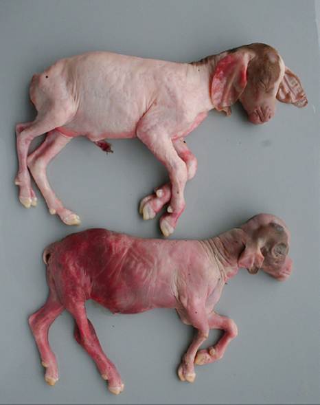
Figure 13.6 Aborted twin Boer kids. The upper fetus was alive at delivery, while the lower fetus had been dead long enough to begin to mummify, as demonstrated by the sunken eye sockets. Although these findings were suggestive of toxoplasmosis, the dam was serologically negative, ruling out toxoplasmosis as the cause of abortion. Source: Courtesy of Dr. M.C. Smith.
requires maintenance of live organisms; many laboratories no longer perform it (Reiter-Owona et al. 1999). ELISA tests, which are easily automated and use soluble antigens from disrupted toxoplasma organisms, have become more popular (Denmark and Chessum 1978; Buxton 1998). Other serologic tests currently used include the indirect hemagglutination test, indirect immunofluorescent antibody test, and latex agglutination test (Buxton 1998; Sudan et al. 2013).
If the diagnosis is to be made by histology, it is very important that placenta be submitted. Small yellowish- white foci of mineralization confined to cotyledons are apparent grossly if abortion is delayed until 45 days or more after infection. Washing the cotyledons thoroughly in isotonic saline solution makes deeper foci easier to visualize. Another useful technique is to compress the cotyledon with a glass microscope slide; the mineralized foci resist squashing. Microscopic foci of necrosis can be identified after 30 days; tachyzooites are sparse in these lesions and difficult to locate (Dubey 1988). Non-suppurative encephalomyelitis is found more consistently than myocarditis, but even under ideal experimental conditions it is difficult to find T. gondii organisms in tissue sections. Very careful histologic examination or immunohistochemistry distinguishes Toxoplasma from the far less common Neospora infection (Dubey et al. 1992). PCR tests might also be employed (Sudan et al. 2013).
Prevention
The control of toxoplasmosis can be approached in several ways. The first is to prevent exposure of susceptible goats to the oocysts in cat feces during the period of danger, which is pregnancy. In particular, grain should be stored in covered containers and the mangers kept clean. Contamination of the hay supply (by cats living in the hay barn) has been implicated in several outbreaks (McSporran et al. 1985; Nurse and Lenghaus 1986). If possible, feed the hay off the top of the stack to the non-pregnant does and young stock. Because exclusion of all cats from a farm is very difficult, it is usually suggested to maintain a stable population of adult neutered cats; kittens younger than 6 months of age are far more apt to shed large numbers of oocysts than are adults. A vasectomized tomcat might be helpful in keeping stray cats off the farm, but this technique has not been evaluated. Raw meat should not be fed to cats. Unfortunately, oocyst shedding has been documented in adult cats after reexposure, especially to heterologous strains (Zulpo 2018), so adult cats do present some risk to naive goats.
Exposure of doelings to a contaminated environment (buildings where feral cats live or pastures spread with manure from such buildings) before breeding to develop protective immunity might be effective (Buxton 1998). Rubbing their noses with placentas from aborted fetuses has been suggested as one way of infecting doelings (Delahaye 1987), but this is ill advised if other causes of abortion such as chlamydiosis are active in the herd. In the milking herd this would increase the risk of acute shedding of toxoplasmosis in milk. Improperly cooked goat meat would also present a higher risk to the consumer, as the goats are apt to be persistently infected.
A live toxoplasmosis vaccine (Toxovax®, MSD Animal Health) available for sheep in Europe and New Zealand might be effective in goats (Chartier and Mallereau 2001), but will probably never be licensed in the United States because it is infective to humans. It cannot be given to pregnant animals and should be given at least 3 weeks before mating. Repeated administration of killed vaccine or vaccination with a related but non-pathogenic organism (Hammondia) might be effective in preventing abortion (Dubey 1981; Munday and Dubey 1988), but would certainly be expensive; such vaccines are unlikely to be marketed. Chemoprophylaxis by feeding ionophores such as monensin during pregnancy, at the normal anticoccidial rate, initially showed promise (Blewett and Trees 1987; Buxton et al. 1988). However, fatal mixing errors may occur with ionophores. Daily decoquinate at 2 mg/kg bodyweight is a safer drug to try and is licensed in the United Kingdom for controlling toxoplasmosis in sheep (Buxton 1998), but extralabel use of drugs in feed is not legal in the United States.
Sulfadimidine (sulfamethazine) at 33 mg/kg bodyweight intramuscularly, four doses, every 48 hours, has shown promise for reducing losses in goat herds experiencing abortion storms due to toxoplasmosis in Greece (Giadinis et al. 2013), but this drug is not available in an injectable form in the United States and other sulfonamides have not been evaluated for this purpose in goats. When abortions caused by toxoplasmosis are diagnosed, emphasis should be put on properly disposing of fetuses and placentas, wearing protective gloves when handling these items, and properly pasteurizing milk and cooking meat. Pregnant women should be especially careful.
Miscellaneous Infections
Other infections that induce a febrile response or generalized illness may cause abortion in does. One such agent is foot and mouth disease, as described in Chapter 4. Theileriosis is described in Chapter 7. Others that lead to occasional abortions are described below.
Bluetongue
Bluetongue is an infectious, non-contagious disease of ruminants, especially sheep, caused by an orbivirus and transmitted by Culicoides midges. The disease occurs in North and South America, Africa, the Middle East, Asia, and recently in northern Europe. Abortion and birth of malformed lambs are common in sheep. Goats are frequently infected in endemic regions, as evidenced by serologic surveys (Lefevre and Calvez 1986; Flanagan et al. 1995; Ting et al. 2005), but clinical signs are rarely recorded (Van Tonder 1975) and bluetongue is unlikely to be the cause of abortions in goats even when they are seropositive. However, bluetongue virus (BTV-1) has been demonstrated in aborted goat fetuses in India (Chauhan et al. 2014) and transplacental infections have been reproduced experimentally by infecting goats on day 61 of pregnancy using BTV-8 (Belbis et al. 2013). See further discussion of bluetongue in Chapter 10.
Border Disease and Bovine Virus Diarrhea
Border disease of sheep is caused by a pestivirus serologically similar to bovine virus diarrhea (BVD) virus (Nettleton et al. 1998), and BVD strains isolated from cattle can also produce disease in small ruminants. Likewise, the border disease virus can cause disease similar to BVD in cattle (Braun et al. 2019). The disease has been underdiagnosed in the past, because early work concentrated on neurologic and fleece changes in lambs infected in utero. As the pathogenesis of the disease became clearer, in conjunction with important discoveries relating to BVD virus, it was learned that lambs infected in utero during the first half of pregnancy often remain persistently infected for life. Some fail to thrive, but others are silent carriers and shedders of the virus and can introduce the disease into other flocks. Infected ewes regularly produce persistently infected lambs. Also, some strains of the virus apparently do not produce neurologic and fleece changes (Bonniwell et al. 1987).
Spontaneous border disease (tremors, no haircoat changes) has been reported only rarely in goats (Loken et al. 1982b; Loken 2000). Of three goats in Norway that seroconverted during pregnancy (out of 145 experiencing gestation failure and being sampled twice), one had an apparent false pregnancy while each of the other two produced a normal and a weak-born kid (Loken 1990). A novel border disease virus was isolated from aborted goat kids in Italy (De Mia et al. 2005). Numerous abortions, stillbirths, and weak but live kids were reported from two herds in Italy from which a genotype 3 border disease virus was isolated. Two kids, 2-3 weeks old, and one yearling goat from this outbreak had virus demonstrated in the skin of the ear, and the yearling was proven to be persistently infected in all tissues but had no gross lesions at necropsy (Rosamilia et al. 2014). Additionally, an experimental contagious ecthyma vaccine contaminated with a non-cytopathic pes- tivirus was associated with reproductive failure in 213 of 261 goats vaccinated early in pregnancy (Loken et al. 1991), but no kids with border disease were born.
A BVD type 2 virus was isolated from goats in a Korean herd experiencing abortions, stillbirths, and neonatal deaths (Kim et al. 2006). Non-pregnant goats exposed on pasture to calves persistently infected with BVD serocon- verted rapidly, with 10 of 10 positive within six weeks (Broaddus et al. 2007). When pregnant goats were penned with persistently infected heifers, 12 of 24 aborted (Broaddus et al. 2009). In another instance, a doe that was penned by chance with a calf persistently infected with BVD after 37 days of gestation gave birth to a persistently infected kid from which virus could be demonstrated in serum, saliva, tears, nasal secretions, and hair (Bachofen et al. 2013). Two of three pregnant does later housed with this animal in early pregnancy had persistently infected kids, and two live but small kids were produced from one of them. Both the original persistently infected goat and the second passage kids developed severe hypoplastic anemia necessitating euthanasia. In a later study, all five pregnant goats exposed before 45 days of gestation to the original infected calf aborted, the more usual outcome of exposure to BVD in early pregnancy.
Border disease has been experimentally reproduced in goats by inoculation of tissues of affected sheep; abortions and ataxic or shaker kids have been produced (Huck 1973; Barlow et al. 1975). In another study, intramuscular inoculation of a cytopathic bovine strain of BVD virus to does approximately 40 days into pregnancy resulted in partial resorption of the fetuses and presence of 1-1.5 L of fluid (hydrometra) in the uterus on slaughter at four months after breeding. Kids were clinically normal when infection occurred at 100 days (Loken 1987). In a later study comparing a border disease strain and a BVD strain, results were similar in both groups, and all goats inoculated around 40 days experienced reproductive failure. At least two viable kids were produced from later inoculations that were persistently infected (Loken and Bjerkas 1991). Some aborted and weak kids had presuckle antibodies. In another study, kids that survived experimental infection during gestation were not persistently infected (Depner et al. 1991). Placental lesions, including multiple pinpoint foci of necrosis in the cotyledons, resemble those of toxoplasmosis (Barlow et al. 1975; Loken 1987). Birthcoat changes have not been reproduced experimentally in goats (Orr and Barlow 1978).
Caprine Herpesvirus
This agent causes a generalized infection in kids and adult goats, but not in lambs or calves; it is discussed in detail in Chapter 12. A diphasic fever has been observed in adults and may contribute to the occurrence of abortion. Fetuses aborted from experimentally infected does were passed in a very autolytic state three to eight weeks after infection in one study (Berrios et al. 1975). In another study, fetal death occurred by four days after inoculation in one goat and the fetuses were harvested by cesarean section the next day, while abortion occurred in the other inoculated doe on day seven without previous ultrasonographic evidence of fetal death. Virus was recovered from the fresh placentas and from one fetal lung, but maternal rather than fetal infection was believed to be the cause of fetal death (Waldvogel et al. 1981). More recently, the caprine herpesvirus has caused cross-placental infection and death of neonates in conjunction with abortion storms. Fetal infection has been confirmed by PCR in aborted fetuses (Keuser et al. 2002; Chenier et al. 2004; Gonzalez et al. 2017) and neonates (Roperto et al. 2000), and foci of coagulative necrosis with intranuclear inclusion bodies may be found in numerous tissues, including the liver, lungs, kidney, and thymus (Williams et al. 1997; Chenier et al. 2004; Uzal et al. 2004; McCoy et al. 2007; Gonzalez et al. 2017). Viral isolation is difficult if the fetus is autolyzed (Tempesta et al. 2004). A marked increase in serologic titer of the dam to the caprine (not bovine) herpesvirus would be strongly suggestive of the diagnosis.
In one follow-up study of a meat goat herd that experienced a caprine herpesvirus abortion storm, there were no reproductive repercussions the following year. Many does that aborted rebred in the same season, and kids born from these does and from does that were infected but did not abort during the initial outbreak remained seronegative 10 months later (McCoy et al. 2007).
Nairobi Sheep Disease
Nairobi sheep disease is a non-contagious tick-transmitted bunyavirus of small ruminants (Terpstra 1990; Committee on Foreign and Emerging Diseases 2008). The virus has been found especially in Kenya, but also in other parts of East and Central Africa. The same virus is found in the Indian subcontinent, where it is known as the Ganjam virus (Baron and Holzer 2015). Signs are more severe in sheep than in goats and include fever, hemorrhagic gastroenteritis, abortion, and increased mortality rates. Histologically, glomerulo-tubular nephritis is present (Mugera and Chema 1967). Tick control and prophylactic vaccination could be recommended, but are not warranted for controlling Nairobi sheep disease in enzootic areas, except when susceptible animals are introduced (Davies and Terpstra 2004). The disease is discussed in Chapter 10.
Peste des Petits Ruminants
Peste des petits ruminants is a morbillivirus infection of goats and sheep in Africa, the Middle East, and Asia. Signs include stomatitis, diarrhea, pneumonia, and a fever that persists for five to eight days. Pregnant animals may abort. The disease is discussed in Chapter 10.
Rift Valley Fever
Rift Valley fever is a mosquito-borne bunyavirus (Phlebovirus genus) disease of ruminants and humans in Africa (Yedloutschnig and Walker 1986; Kasari et al. 2008) and is described in detail in Chapter 11. Abortion may come after fever and viremia in adults (Daubney et al. 1931). Goats are generally more resistant than sheep or cattle (Erasmus and Coetzer 1981) and show a lower abortion rate. Very young kids also may die. Hemorrhages and hepatic necrosis are found at necropsy. Because serious human infection also occurs, abortion samples should be collected carefully. A modified live virus vaccine produces lifelong immunity, but must not be given to early-pregnant sheep or goats, because it produces fetal malformations, especially of the brain (Bath and de Wet 2000). Newer, safer vaccines are under development (Kortekaas 2014).
Wesselsbron Disease
Wesselsbron disease is a mosquito-borne flavivirus disease of sheep, cattle, and goats. It is restricted to sub-Saharan Africa and may cause abortion in pregnant does (Van Tonder 1975; Van der Westhuysen et al. 1988) and ewes and neonatal mortality. Seroconversion of the aborting doe can be used for diagnosis (Mushi et al. 1998). Adult nonpregnant does rarely show any signs except fever. Differentiation from Rift Valley fever is described in Chapter 11. Live virus vaccines against both of these diseases can cause abortion if given during pregnancy. A low incidence of congenital anomalies (i.e., arthrogryposis, porencephaly, hydranencephaly, cerebellar hypoplasia) is reported in sheep infected with Wesselsbron disease, but apparently not documented in goats.
Mycoplasmosis
Numerous Mycoplasma species have been isolated from goats and associated with specific clinical syndromes discussed elsewhere in this text (i.e., arthritis, keratoconjunctivitis, mastitis, pneumonia). Abortions occasionally occur during outbreaks of mycoplasma-induced disease, but documentation that mycoplasms caused these abortions is often lacking. For instance, an abortion rate of 80% was reported in a pen of 50 goats during an outbreak of mastitis and arthritis caused by Mycoplasma putrefaciens (DaMassa et al. 1987). In another outbreak, however, transplacental infection was documented by the birth of weak kids already demonstrating swollen joints from which Mycoplasma mycoides subsp. mycoides was isolated (Bar-Moshe and Rapapport 1981) and M. mycoides subsp. mycoides large colony was isolated from two aborted kids in Spain (Rodriguez et al. 1995). This organism has been reclassified as M. mycoides subsp. capri (Manso-Silvan et al. 2009).
Yersiniosis
Yersinia pseudotuberculosis is a zoonotic bacterium commonly carried by wild birds or rodents. Fecal-oral infection of goats can result in establishment of an enteric infection with subsequent bacteremia. Abortions and early neonatal deaths of kids have been reported (Sulochana and Sudharma 1985; Witte et al. 1985; Albert 1988; Giannitti et al. 2014). Opaque white foci were seen in cotyledons, and microscopic lesions included suppurative placentitis and suppurative fetal pneumonia (Witte et al. 1985; Giannitti et al. 2014). Tetracycline may be useful for halting an abortion storm. Yersiniosis is discussed further in Chapter 10.
Tick-Borne Fever
Anaplasma phagocytophilum (formerly Ehrlichia, Cytoecetes, Rickettsia phagocytophila) causes a mild to acute, non- contagious disease of sheep, goats, and cattle in the United Kingdom, Europe, Africa, and India (Baas 1986). Although the organism is considered to be a common tick-borne zoonotic agent in the United States, reports of clinical disease in goats in the country are lacking. Natural transmission requires ticks such as Ixodes ricinus or Rhipicephalus haemaphysaloides, but the disease can be spread by anything that transfers blood. Signs include fever, listlessness, decreased milk production, lameness, and abortion. Secondary infections (subsequent to neutropenia) with Staphylococcus, Pasteurella, Listeria, and other organisms occur commonly and may contribute to abortion.
Diagnosis is by demonstration of cytoplasmic inclusions in granulocytes and monocytes in the blood and by serologic or polymerase chain reaction tests. The inclusions are grayish-blue in Giemsa-stained blood smears, which should be made as soon after abortion as possible. The organism does not invade the fetus, and thus smears of fetal tissue are not helpful (Scott 1983). Tetracycline is used for treatment (Anika et al. 1986). Pregnant animals should not be placed on pastures heavily infested with ticks. Pasture exposure of kids may prevent later economic losses from decreased production of milking goats (Melby 1984).
Anaplasmosis
Anaplasma ovis infection in goats is widespread (Renneker et al. 2013) and usually causes a subclinical, mild, febrile disease unless the goats are stressed. However, Boer goats in semiarid regions of South Africa that had to walk long distances for food aborted and aborting does had low hematocrits. Organisms were found in the red blood cells of these goats. Of 20 experimentally infected pregnant goats, 10 aborted or resorbed their fetuses under conditions that included driving them 800 m twice a day (Barry and van Niekerk 1990). Goats showed respiratory distress and pulmonary edema when driven. The average peak in body temperature was 1.1 °C (2 °F) above preinfection levels and the mean hematocrit dropped from 30 to 22% at the peak of parasitemia, with the lowest recorded hematocrit being 10%. Transplacental infection occurred; A. ovis organisms were observed in red blood cells of aborted and live-born kids. Means of control of this tick-borne infection, which is usually subclinical, are yet to be determined. The disease is discussed further in Chapter 7.
Neosporosis
Neospora caninum is a protozoal parasite similar to T. gondii that can cause abortion in goats. Cattle are more commonly affected. Whitetail deer may be involved in a sylvatic cycle and dogs and coyotes have been identified as definitive hosts (Gay 2006). Transplacental infections have been documented in goats (Mesquita et al. 2013) and vertical transmission from the dam may be a common route of infection.
Experimental infection of Pygmy goats has produced resorption, abortion, and stillbirths (Lindsay et al. 1995). Diagnosis is by isolation from the placenta or by histology and immunohistochemistry, which distinguishes Neospora from Toxoplasma infection (Dubey et al. 1992). Dual infections with both parasites have been identified (Anastasia et al. 2013). Lesions in the fetal brain include meningoencephalitis with necrosis, gliosis, perivascular cuffs, and protozoal tissue cysts approximately 10 μm in diameter (Dubey et al. 1996). Hydrocephalus and cerebellar atrophy were seen in this case. Diagnosis by PCR is also possible (Eleni et al. 2004).
Many serologic surveys have been done in the last 20 years in Europe, South America, Africa, and Asia, with variable results. The prevalence of antibodies was only 1-2% in dairy goats in western France (Chartier et al. 2000). In a survey of 50 mixed sheep and goat flocks in Greece, 7% of 375 goats were seropositive for Neospora caninum (Anastasia et al. 2013). Seroprevalence in 384 dairy goats in Bahia State, Brazil, was 15% (Uzeda et al. 2007). Seroprevalence in goats is probably affected by the prevalence of dogs on goat farms. Presence of antibodies in the dam does not prove cause of abortion.
Sarcocystosis
Sarcocystis is a genus of cyst-forming protozoa. Cysts are frequently noted as an incidental finding on histologic examination of cardiac and skeletal muscle of many species. Some Sarcocystis species cause macroscopic lesions that may result in carcass condemnation. In a New Zealand study, sarcocysts with mean cross-sectional diameters of 69 ? 54 μm were found in 28% of samples from diaphragms of feral goats. When muscle from goat carcasses was fed to dogs and cats, only the dogs shed sporocysts in their feces (Collins and Crawford 1978). A slaughterhouse study from Brazil demonstrated microscopic cysts (thought to be Sarcocystis capracanis) in 92% of 120 meat goat carcasses (Bittencourt et al. 2016). Experimental inoculation of goats 75-105 days' pregnant with 10 000 sporocysts from coyote feces and believed to be S. capracanis resulted in illness and abortions. Because no Sarcocystis meronts were found in the placenta or fetuses, the abortions were probably an indirect result of acute sarcocystosis. Naturally occurring abortion caused by sarcocystosis has been reported once in goats (Mackie et al. 1992; Mackie and Dubey 1996).