Microscopy, Cytology, and Histopathology
Microscopic examination of clinical specimens using wet smears, cytopathology, and histopathology are cornerstone diagnostic tools for blastomycosis in humans and animals (Saccente and Woods 2010).
The classic appearance is of large (8-15 μm), round, multinucleate yeast-like cells with thick, double refractile walls that may have a single bud with broad bases (Guarner and Brandt 2011) (Fig. 8.3). Small yeast forms are also known to occur and may be difficult to distinguish histologically from the yeast-like cells of other dimorphic fungi (Guarner and Brandt 2011).The most expedient test is a wet preparation using potassium hydroxide (KOH) solution, with or without staining with calcofluor white or lactophenol blue (Saccente and Woods 2010). The sensitivity of this test is poor: a study found the yield of sputum for KOH preparation in human pulmonary blastomycosis to be 25% for a single specimen (Martynowicz and Prakash 2002). Nonetheless, examination of multiple specimens often leads to the correct diagnosis. For example, in a series of 100 persons with pulmonary blastomycosis, KOH preparation of respiratory samples (invasively and noninvasively collected) led to the immediate diagnosis in 66% of cases (Patel et al. 1999).
Visualization of Blastomyces yeast-like cells is enhanced with cytologic and histopathologic preparations. Cytology stains like Papanicolaou and Wright stains can be used to identify the organism in respiratory specimens (usually sputum and bronchoalveolar lavage in humans and transtracheal lavage or transthoracic fine- needle aspirates in dogs), lymph node aspirates, and impression smears or discharge from cutaneous lesions (Martynowicz and Prakash 2002). In tissue, the yeast-like
Fig. 8.3 Modified Wright's stain of impression smears collected from a hilar mass during necropsy of a 4-year- old mixed breed male dog, viewed at 60 x.
Note the large, round yeast-like cells with thick, double refractile walls and broad-based budding (Courtesy of Dr. Angelica Galezowski, DVM, MVetSc, DACVP, Faculty of Veterinary Medicine, University of Calgary, Canada)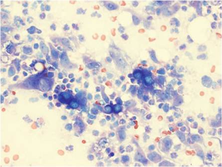
cells may first be visualized with hematoxylin and eosin, although sensitivity and specificity are greatly improved by use of fungal stains like periodic acid-Schiff (PAS) or Grocott-Gomori methenamine-silver nitrate (GMS) (Saccente and Woods 2010). The host response is characterized by mixed inflammatory reaction, predominated first by neutrophils and eventually by noncaseating granulomas (Saccente and Woods 2010).
Cytologic and histopathologic examinations are frequently used in medical and veterinary practice. These tests perform well in diagnosing blastomycosis compared to culture (Patel et al. 2010), and with faster turnaround time. Cytologic examination led to the diagnosis in 71% of cases of canine disease from Louisiana, and histopathologic examination diagnosed an additional 9% (Arceneaux et al. 1998).
8.4.3