Neoplastic diseases of the small intestines
Carolyn J. Henry
Introduction
Small intestinal (SI) neoplasia is uncommon, noted in 0.3% and 0.7% of all canine and feline necropsy submissions, respectively, each year.1 Of the non-lymphoid SI tumors (see 9.3 for lymphoid tumors), carcinomas predominate in both species.2 Leiomyosarcomas comprise the second most commonly reported non-lymphoid gastrointestinal malignancy in dogs.2- 4 Other reported canine SI tumors include leiomyomas, fibrosarcomas, undifferentiated sarcomas, mast cell tumors (MCT), carcinoids (see 9.4.6), neurilemomas, and extramedullary plasmacytomas.1,2,5-7 Although uncommon, leiomyosarcomas have been reported in cats, as have MCTs, hemangiosarcomas, adenomatous polyps, and extraskeletal osteosarcomas.8-16
Incidence
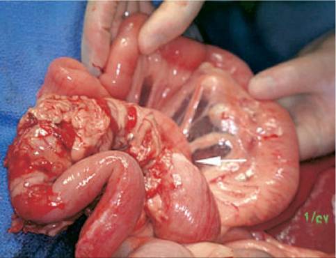
Figure 5.10:
Intestinal adenocarcinoma.
This figure shows an adenocarcinoma located in the ileum of a dog. The mesentery is adhered to the affected portions of intestine. The lesion is tubular in nature and causes annular narrowing of the bowel noted by a white band of constriction (arrow).Although the small intestine comprises 90% of the length of the intestinal tract, most canine non-lymphoid intestinal tumors, other than MCTs, are located in the colon or rectum, not in the more proximal segments.1,5,17 This is true in human beings as well, where small bowel tumors account for only 3% of all GI malignancies.18 In contrast, previous reports have indicated that 90% of feline intestinal tumors occur in the small intestines.1 However, more recent data suggest that this reported site predilection in cats may no longer hold true (personal unpublished data). Multiple hypotheses have been proposed to explain the low incidence of SI tumors in dogs and humans.
Proposed factors include the rapid transit time of potential carcinogens in the proximal segments compared to the colon, an inability of the SI flora to transform procarcinogens into their active metabolites, the presence of microsomal enzymes in the small intestine that detoxify carcinogens, and local immunosurveillance by IgA-secreting lymphocytes and B cells in the distal ileum.19 However, the reason for the previously reported SI site predilection in cats remains unexplained. A role for retroviral diseases in the etiology of feline non-lymphoid intestinal cancer has not been identified.1,2,20 Any segment of the canine SI may be affected, although sarcomas occur more often in the jejunum than in other segments. The jejunum and ileum are affected more often in cats than is the duodenum.1,11,20,21Small intestinal neoplasia is generally a disease of older animals, occurring at a mean age of approximately 9 years in dogs and a mean and median age of 8.7 and 11 years, respectively, in cats.2,3,11,20 Some tumor types, especially leiomyosarcomas, have been reported in very young animals.22 Accordingly, age should not be used to exclude a diagnosis of SI neoplasia. No clear breed predispositions have been identified for canine SI cancer except with MCT, for which miniature breeds, especially the Maltese, are at increased risk.5 Siamese cats appear to be predisposed to intestinal cancer.2,11,20,21 Conflicting views exist regarding gender predispositions for intestinal cancer in both species, with some reports indicating a male predisposition and others noting a female predisposition.1-3,21,22 In humans, there is a slight male predominance for malignant small bowel tumors.19
The most common primary SI tumor in both species, with the exception of lymphoma, are those of epithelial origin, the majority of which are malignant.1 Of the four types of malignant epithelial tumors (adenocarcinoma, mucinous adenocarcinoma, signet ring cell carcinoma, and undifferentiated or solid carcinoma) adenocarcinomas predominate.
The tumors often cause annular constriction (Figure 5.10) and may become quite large before they are clinically detectable. Feline intestinal adenocarcinomas (ACA) have been histologically subtyped as tubular, undifferentiated, or mucinous.11,20,23 Of these, mucinous ACA are the least common.23 Approximately one third of feline intestinal ACAs have areas of osseous or cartilaginous metaplasia.1 Tumor grading based on degree of cellular differentiation may not be clinically relevant. In a series of cats with intestinal ACAs, papillary ACAs were described as the most differentiated tumors, yet they were associated with the highest rate of metastasis.21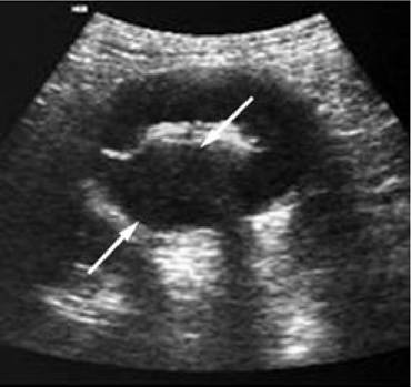
Figure 5.11:
Ultrasonographic appearance of a small intestinal mass. Transverse sonogram of a hypoechoic circumferential mural lesion in the small intestine. The hyperechoic central portion represents the bowel lumen and arrows indicate the extent of wall thickening due to neoplasia.
Leiomyomas and leiomyosarcomas originate from smooth muscle and are the most common mesenchymal tumors affecting the canine GI tract.2-4-24 Over 50% of dogs with intestinal leiomyosarcoma have intra-abdominal metastasis at the time of diagnosis, thus emphasizing the need for presurgical tumor staging.25 Metastatic disease may occur through hematogenous or lymphatic routes, as well as by transcoelomic spread and tumor seeding.1 Metastatic lesions are occasionally found in the testes, although the route by which this occurs is unclear.7,26
Clinical signs
Weight loss, vomiting, and anorexia are the most common presenting complaints associated with SI cancer in dogs and cats.2,3,20,23 In dogs, weight loss and vomiting are more common with duodenal or jejunal lesions whereas in cats, there is equal site distribution for masses associated with these presenting complaints.2 Diarrhea and tenesmus are more typical for ileal or colonic masses than for proximal SI lesions.2 Other signs reported with SI cancer include melena, abdominal distension, and lethargy.2,3,20,25,27 Paraneoplastic syndromes have been reported with smooth muscle tumors and include hypoglycemia (leiomyoma and leiomyosarcoma) and nephrogenic diabetes insipidus (leiomyosarcoma).25,27,28 Dogs with paraneoplastic hypoglycemia may present with CNS signs including seizures and ataxia.25,29,31 Hypoglycemia may also occur secondary to tumor-related peritonitis, especially with smooth muscle tumors, which are prone to grow quite large and can subsequently rupture.25 Polyuria and polydipsia (PU/ PD) were noted in over one third of the dogs with GI leiomyosarcoma in one report and have been described in other reports of smooth muscle tumors.25,28,31 In one case, the cause of PU/PD was apparently tumor-related nephrogenic diabetes insipidus; however, the cause has remained undetermined in others.25,28,31
Diagnostics
Physical examination findings may aid in the diagnosis, as a palpable mass is found in approximately half of all cases.2,3,20 Plain radiographs are generally less sensitive than palpation or abdominal ultrasound for detection of intestinal masses, especially in cats.2,20 However, plain radiographs were sufficient to warrant surgical exploration in 65% of cats in one report.23 Contrast radiography may enhance tumor visualization and was diagnostic for tumor-related obstruction in 13 of 15 cats in one study.20 Abdominal ultrasound is becoming the gold standard for imaging intestinal neoplasia as it permits more accurate assessment of intestinal wall layering and facilitates the differentiation of neoplasia from nonspecific enteritis.32 Abdominal ultrasound did permit identification of intestinal neoplasia in 87% to 90% of cases in two reported case series.3,32 Dogs in which intestinal wall layers are no longer visible (Figure 5.11) are reportedly over 50 times more likely to have intestinal neoplasia than nonspecific enteritis.32 Segmental intestinal wall thickening of mixed echogenicity in cats supports a diagnosis of ACA over that of lymphoma.33 However, definitive diagnosis is generally made by fine needle aspiration of a palpable or ultrasound-imaged mass or by histopathological examination of a tissue biopsy collected during laparoscopy or exploratory laparotomy.
Preoperative lab work should include a CBC, serum chemistry profile, and urinalysis.
Anemia and leukocytosis are the predominant CBC abnormalities noted in both species.2,20 The leukocytosis is generally characterized by neutrophilia.2 Lymphopenia was noted in 7 of 11 affected cats in one re- port.23 Decreased neutrophil numbers, a degenerative left shift, or toxic changes in neutrophils should alert the clinician to the likelihood of tumor rupture and peritonitis, and is commonly reported with leiomyosarcomas.25 Biochemical abnormalities in dogs may include hypoproteinemia due to protein loss or decreased production, and hypoglycemia occurring either as a paraneoplastic syndrome or secondary to sepsis or hepatic failure.3,25 In cats, hypoproteinemia, hyperglycemia, azotemia, hypercholesterolemia, and increased activities of alanine transaminase and alkaline phosphatase have been re- ported.2,20,23Staging
Preoperative staging procedures should include a thorough physical examination to assess for unusual sites of metastasis, three-view thoracic radiographs, and an abdominal ultrasound. The mesenteric lymph nodes are the most frequent site of intestinal carcinoma metastasis.2,7,23 The rate of intra-abdomi- nal metastasis to lymph nodes, liver, and mesentery exceeds 58% in dogs and 70% in cats with intestinal ACA.3,7,20 Metastatic rates are slightly lower with leiomyosarcomas, ranging from 18% to 54%.3,25,27 Lung metastasis is an uncommon feature with any of the different types of intestinal neopla- sia.3,7,20,23,27 Metastases to unusual sites including the testes and skin may occur with intestinal ACA, thus emphasizing the need for a thorough physical examination.7,26,34
Treatment
Surgery is the only proven beneficial treatment for non-lym- phoid SI cancer.3,35 Conversely, surgery is generally unrewarding for visceral MCT in dogs, with a reported median survival time (MST) of 18 days.36 The goals of exploratory laparotomy when SI neoplasia is expected are: 1) to facilitate complete excision of an identified mass and 2) to visualize and biopsy affected organs, so that a definitive diagnosis and staging can be achieved.
Surgical margins should ideally include 4 to 8 cm of normal appearing tissue.37 Complete abdominal exploration should be performed to assess for metastatic disease. Also, lymph nodes draining the affected site should be biopsied. In general, the duodenum is drained by the hepatic and pancreaticoduodenal nodes, the jejunum is drained by the jejunal lymph nodes found at the origin of the jejunal vessels, and the ileum is drained by the jejunal and colic nodes.38 Although metastases may have a negative impact on overall outcome, several reports caution against recommending euthanasia at the time of surgery on the basis of presence of metastatic disease alone. This is especially true for dogs with leiomyosarcoma, in which survival times exceeding three years have been reported, even in cases with metastatic disease.27 For patients with ACA and metastasis, a one-year survival rate of 20% has been reported for dogs and a mean survival time of one year has been reported for cats.3,20 Tissues should be submitted for histopathological evaluation and, when bowel rupture has occurred, bacterial culture and sensitivity testing is indicated.Immunohistochemistry (IHC) may be needed in order to reach a definitive diagnosis in some cases. The IHC markers of value for intestinal masses include cytokeratin for epithelial tumors; vimentin, desmin, and α-smooth muscle actin for spindle cell tumors; and c-kit and mast cell tryptase for MCTs.17,39,40
The roles of chemotherapy and radiation therapy have not been defined for non-lymphoid SI cancer in dogs and cats.3,35 When chemotherapy has been attempted for visceral MCT in dogs, the results have been poor.36
Prognosis
Prognosis for canine SI cancer is largely related to tumor type. Amongst the non-lymphoid malignancies, leiomyosarcomas are associated with the most favorable prognosis, with a reported MST from 12 to 21.3 months for dogs surviving the immediate post-operative period.25,30 In one report, female gender was a poor prognostic factor for canine intestinal ACA.
Female dogs had a MST of 28 days, compared to 233 days for males.41 This finding has not been duplicated in other reports. Dogs with intestinal ACA survive an average of 10 months, whereas average post-surgical survival time in cats has been reported to only be 2.5 to 5 months.3,11,23 However, a more favorable outcome has been reported for cats with tubular ACA (11 months), than for those with mucinous and undifferentiated ACA (4 months).23 Metastatic disease is an important prognostic factor for dogs with intestinal ACA. In one report, dogs with metastatic disease had a three-month MST and a 20% one-year survival rate, compared to a fifteen-month MST and 66.7% one-year survival rate for those without metastases.3 In cats with ACA, metastatic disease has not been reported to have a similarly profound affect on prognosis. Although one report indicated a five-month mean survival time in cats with metastasis compared to ten months in cats without metastasis, another series reported that five cats with lymph node metastasis had a mean survival time of twelve months.20 This disparity in the literature and the fact that survival beyond two years has been reported in a cat with carcinomatosis and visceral metastasis indicates that metastatic disease should not be used as a sole predictor of prognosis.20 Prognosis is less controversial for visceral MCT in dogs, for which the reported MST is 18 days.36Non-malignant lesions, including polyps and leiomyomas are curable with surgery. In one series of cats with duodenal polyps, surgery provided a complete resolution of signs in 13 of 15 cats and none were reported to have a recurrence.14 Similarly favorable outcomes have been reported for dogs with leiomyomas.31,42
??S> Key Facts
■ The majority of feline intestinal tumors occur in the small bowel, whereas most non-lymphoid intestinal tumors in dogs affect the colon or rectum.
■ Dogs with intestinal smooth muscle tumors may present for signs related to hypoglycemia, rather than for obvious gastrointestinal signs.
■ Ultrasonography is recommended for the identification of intestinal masses and may permit differentiation of neoplasia from nonspecific enteritis.
■ Canine intestinal mast cell tumor warrants a poor prognosis and generally affects miniature breeds, especially the Maltese.
■ The presence of metastatic disease in a cat with intestinal adenocarcinoma does not necessarily warrant a grave prognosis, as survival times of 1 to 2 years after surgical resection have been reported.