ORAL SQUAMOUS CELL CARCINOMA IN DOGS
Background
The second most common oral malignancy in dogs is squamous cell carcinoma. It is usually found in the gingival tissue, although the tongue and tonsils are frequently affected.
Tumors of the tonsil behave very differently from other squamous cell carcinomas and are considered separately in this chapter.Squamous cell carcinoma usually occurs in older dogs; the average age is 9 years. There is no apparent breed or gender predilection, although one study of squamous cell carcinoma of the tongue found that 43% of affected dogs had a white hair coat and 30% were poodles. Papillary squamous cell carcinoma occurs in dogs as young as 2 months of age and is a progressive disease with a high rate of lytic bone involvement. Dogs with this type of tumor are almost always less than 2 years of age (Figure 11-1).
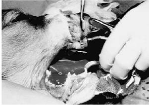
Figure 11-1 An intraoperative biopsy of a 7-week- old puppy with dramatic osteolysis of the underlying mandible due to an oral papillary squamous cell carcinoma. Radiation therapy resulted in a cure. She died 13 years later of unrelated causes. Oral papillary squamous cell carcinoma is a curable disease by either surgery or radiation therapy.
Clinical Parameters
Most oral squamous cell carcinomas occur within the rostral portion of the mouth, and the majority occur in the maxilla. However, in one study the most common area for these tumors was the tonsils. Affected dogs show the same signs as do dogs with other oral tumors. The most common signs are drooling, halitosis, and (occasionally) dysphagia. Most dogs have shown signs for 3 months or less, but some dogs may show signs for 6 months to 1 year before diagnosis.
Clinical Work-up
Biopsy is required to differentiate squamous cell carcinoma from amelanotic melanoma, ulcerated fibrosarcoma, or other less common tumors.
Staging should be performed as previously described for other oral tumors. Squamous cell carcinoma is highly invasive, and high-detail radiographs of the skull often reveal extensive bony lysis. Bony lysis alone should not be relied on for surgical margins or radiotherapy field size, because this underestimates tumor size. CT or MRI scanning is often helpful in planning radiation therapy and/or surgical excision.Gingival squamous cell carcinoma rarely metastasizes. Regional lymph nodes are frequently enlarged at diagnosis, and, as for all oral tumors, they should be aspirated and/or biopsy should be performed on them (even if they are not enlarged) for cytologic or histopathologic examination; however, these nodes are usually reactive. In one study 11 of 33 dogs had lymphadenopathy, and only 3 of these dogs had metastatic disease. After therapy, regional lymph node metastasis is still uncommon; however, metastasis was documented in 5 (21%) of 24 dogs in three case series. In contrast, squamous cell carcinoma of the tongue seems more aggressive; 9 (43%) of 21 dogs in one study developed metastasis to lymph nodes, lung, or bone.
Prognostic Factors
One study reported that dogs with maxillary tumors had a longer average response to radiation therapy (12 months) than did dogs with mandibular (3.4 months) or soft tissue tumors (1.8 months). Eight dogs that were younger than 6 years of age lived for a median of 58 months after radiation; older dogs lived for a median of 6 months. Dogs with rostrally located tumors live longer than dogs with caudal tumors, whereas dogs with tumors that extend both rostrally and caudally have significantly shorter survival times. Therefore larger tumors with larger radiation fields are associated with shorter survival. Megavoltage radiation therapy in dogs with nontonsillar squamous cell carcinoma was associated with shorter median survival in dogs older than 9 years of age compared with younger dogs (median survival, 315 versus 1080 days, respectively).
Therapeutic Approach
Rostral gingival squamous cell carcinoma has a generally low metastatic rate, which makes this malignancy a good candidate for local therapies such as surgery and radiation. Aggressive surgery is necessary to obtain adequate surgical margins. Maxillectomy and mandibulectomy have been used to treat this tumor. From several different reports, median survival ranged from 9 to 18 months. Recurrence was more frequent than metastasis after surgery. Incomplete surgical resection is commonly associated with recurrence, which emphasizes the importance of early diagnosis and obtaining wide surgical margins by mandibulectomy or maxillectomy at the first surgery. Adjunctive radiation therapy may also be useful.
In 33 dogs, orthovoltage radiation therapy without surgery was used to treat oral squamous cell carcinoma to a total dose of approximately 40 Gy. Overall average survival was approximately 14 months; however, the size and location of the tumor, as well as the age of dog, influenced these figures. Recurrence was noted in 15 dogs, metastasis in 3, and serious complications (e.g., bone necrosis) from radiation in 2 dogs. In another study 39 dogs with squamous cell carcinoma were treated with 48 Gy of 60Co teletherapy delivered over 4 weeks on an alternate-day basis. Twelve dogs had local recurrence, 1 dog developed metastases to regional lymph nodes, and 2 dogs had distant metastases. Dogs with rostrally located tumors and dogs with smaller tumors had longer remissions. Median progression-free survival was approximately 13.6 months. In another study dogs with nontonsillar squamous cell carcinoma had a median survival of 450 days after megavoltage radiation therapy.
Surgery combined with radiation gives the best control for gingival squamous cell carcinoma, and postsurgical radiation should be considered for dogs with large tumors or for dogs with tumors that have tumor-present margins on surgical histopathologic examination.
One dog treated in this way had no evidence of disease 16 months after treatment.Squamous cell carcinoma of the tongue is a more aggressive tumor than gingival squamous cell carcinoma. Metastatic disease in this location often determines survival. Unless wide surgical margins are obtained, recurrence is common. Complete removal of the tongue is indicated in some cases and, surprisingly, dogs adapt to this well. In one study five dogs with small tumors were treated with surgery alone and had a median survival time of 8 months. Three of these dogs had local recurrence.
Recurrence of tongue tumors is also common after radiation therapy. Larger tumors in 10 dogs were treated with radiation therapy, and the dogs survived for a median of 4 months; 9 of 10 dogs had recurrence. The 1 dog that received radiation after surgery survived 26 months with no recurrence.
A combined modality approach for larger tumors of the tongue seems warranted for local control, although metastasis occurs in approximately 50% of the cases. Chemotherapy, with or without radiation, should be considered for these tumors as an adjuvant to surgery. Chemotherapeutic agents that have been reported to be of some help in these cases include cisplatin, carboplatin, mitoxantrone, and piroxicam.
Papillary squamous cell carcinoma in young dogs is an aggressive disease. Conservative surgery alone is unlikely to be curative because of the high rate of bone involvement and the young age of the patient. Radiation therapy has a good success rate, although disruption of normal bone growth in young dogs may produce facial malformations. Radiation therapy to a total dose of 40 Gy was used to treat three puppies with this disease.There was no evidence of disease in any dog 10, 32, and 39 months after treatment.
Chemotherapy is rarely indicated for rostral gingival squamous cell carcinoma in dogs due to the low metastatic rate for this tumor. Chemotherapy may be indicated for squamous cell carcinoma of the tongue, tonsil, and caudal location of the mouth because of the high metastatic rate of such tumors.
Subcutaneous bleomycin treatment, as well as doxorubicin, cyclophosphamide, and chlorambucil treatment, failed to induce responses in two dogs with oral squamous cell carcinoma. Cisplatin has caused responses in dogs with metastatic subungual squamous cell carcinoma; however, no responses occurred in five dogs with oral squamous cell carcinoma (including one tongue and one tonsillar tumor). In another report, cisplatin caused a partial response in three of five dogs with oral squamous cell carcinoma for 2, 10, and 15 weeks, respectively. Cisplatin or carboplatin has yet to be fully evaluated for oral squamous cell carcinoma; however, it may be useful in combination therapy for this disease. Mitoxantrone (5 to 6 mg/m2 every 3 weeks) caused responses in four (45%) of nine dogs with squamous cell carcinoma of various sites, including the oral cavity, for between 6 and 21 weeks. Mitoxantrone chemotherapy in combination with surgery was effective in controlling squamous cell carcinoma of the tongue in dogs in another study.Oral piroxicam (0.14 mg/lb once daily) may be helpful in alleviating clinical symptoms and in tumor control.Toxicities associated with the use of this drug include GI and renal. A biochemical profile should be performed before the use of this drug, and if clinical signs associated with GI upset occur, the drug should be discontinued.