ORAL SQUAMOUS CELL CARCINOMA IN CATS
Background
Neoplasia of the oral cavity represents 10% of all tumors diagnosed in cats, and approximately 90% of these are malignant. Squamous cell carcinoma is the most common oral tumor in cats (Figure 11-2).This tumor accounts for approximately 60% of all oral tumors.
The average age of a cat with this disease is 11 to 12 years of age; however, cats as young as 3 years of age may be affected. No gender or breed predilection has been noted.Most oral squamous cell carcinomas in cats occur at the base of the tongue and involve the frenulum (Figure 11-3). This area should be routinely examined in older cats, especially when they are anesthetized for dental or any other procedure that requires sedation or anesthesia. Some speculation has led to the thought that extensive grooming habits of the cat possibly cause the species to contact carcinogens on its hair coat, thereby predisposing the tongue to development of neoplasia.
Clinical Parameters
Cats with oral squamous cell carcinoma most commonly present for a mass or facial asymmetry. Squamous cell carcinomas are characterized by mucosal ulceration, necrosis, and severe suppurative inflammation. Cats with this tumor may pre-
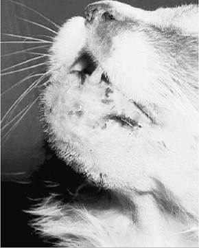
FIGURE 11-2 Squamous cell carcinoma is the most common oral tumor in the cat and one of the three most common oral tumors in the dog. Bone involvement can suggest a more favorable outcome. This 12-year-old spayed female cat had a mandibulectomy, followed by radiation therapy and mitoxantrone chemotherapy. An esophageal feeding tube was placed during this therapy and removed 2 weeks after radiation therapy was discontinued. Her tumor was controlled for 18 months, when metastases were identified. Quality of life was excellent, according to her caregivers.
sent with dysphagia, halitosis, anorexia, nasal discharge, sneezing, pawing at the mouth, changes in eating habits, oral hypersensitivity, loose teeth, weight loss, and drooling of ropelike saliva.
In some cats a small mass is found initially in conjunction with a thorough oral examination during anesthesia for routine dentistry. There may be no symptoms whatsoever at this early stage. In the early stages, differentiating squamous cell carcinoma from gingival proliferation and dental disease may be difficult, so a biopsy should be performed on any oral mass in an older cat, even in absence of the above symptoms.The mandible and maxilla are equally distributed as far as frequency of site that is affected. Bone invasion is common with gingival tumors. Some squamous cell carcinomas arise primarily within the mandible, causing enlargement of the jaw as a result of bony proliferation.These tumors should be differentiated from deep-seated osteomyelitis
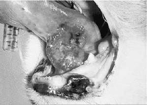
FIGURE 11-3 Sublingual squamous cell carcinoma is a difficult tumor to treat; however, most patients can be made more comfortable with good analgesia and nutritional support, such as assisted tube feeding. Piroxicam (Feldene) is a nonsteroidal antiinflammatory agent and potent analgesic and has been reported to have anticancer effects against selected tumor types. Although its efficacy against sublingual squamous cell carcinoma has yet to be documented in the dog and cat, it is one treatment option for patients that have normal renal function.
generated by severe dental disease and from other malignant tumors, such as osteosarcoma.
Clinical Work-up
Staging of any cat with an oral tumor should be performed as previously described in the dog. High-detail radiography of the skull provides information on bony lysis caused by gingival tumors. As stated previously, however, radiographic appearance of lysis does not occur until more than 50% of the bone has been demineralized; therefore radiography is a poor indicator of tumor margins. Biopsy is required for definitive diagnosis and should be considered in any old cat with severe gingival disease.
Common differential diagnoses include eosinophilic granuloma and fibrosarcoma.Metastasis is rare, although mandibular lymph nodes may be involved and should be evaluated by cytologic or histopathologic examination. Although lymphadenopathy is usually assumed to signal possible metastatic disease, two studies found that less than 50% of cats with enlarged lymph nodes had histologic evidence of metastatic disease. Lymph node metastasis was seen in 8 of 59 cats in these two studies. Metastatic disease may occur late in the course of the disease, or perhaps metastases are slow to progress.This is further supported by 1 cat that had no evidence of metastatic progression 16 months after lymph node metastasis was detected.
Therapeutic Approach
In five cats with squamous cell carcinoma of the mandible, resection alone was not very successful in maintaining a remission. Recurrence occurred in four cats within 5 to 12 months of surgery despite aggressive surgical technique. These tumors were all large (between 2 and 4 cm in diameter), and all invaded bone. In another study, surgery alone resulted in a median survival of 6 weeks for seven cats. Small tumors, particularly those located rostrally on the mandible, may be more amenable to complete surgical excision.
Radiation therapy used alone for the treatment of this disease has also not been rewarding. Radiation therapy (orthovoltage, 52 Gy) was used to treat 11 cats with oral squamous cell carcinoma. Treatment included ethanidazole, which is a hypoxic cell sensitizer that was injected intratu- morally. Eight cats were evaluated for response. Four died from complications of therapy, which included tissue necrosis and ischemia of the tongue, between 45 and 341 days after radiation (median, 114 days). The other 4 cats had tumor recurrence at 125 to 331 days after radiation (median, 170 days). No cats were alive 1 year after treatment. Overall median survival was 132 days. In another study, radiation alone or in combination with chemotherapy or with hyperthermia resulted in a median survival of 10 weeks for 45 cats.
Combination treatment, with mandibulectomy followed by external beam radiation to a dose of 40 Gy to 45 Gy starting 10 to 15 days after surgery, has demonstrated more success. Six of seven cats had tumor recurrence between 3 and 36 months after treatment (median, 12.5 months). One cat died but showed no evidence of disease 14 months after completing radiation therapy.
Placement of a gastrostomy tube by endoscopy or an esophagostomy tube at the time of surgery may prolong survival and definitely facilitates nutritional supplementation in cats undergoing surgery or radiation therapy for oral squamous cell carcinoma (Figure 11-4). The tube allows enteral feeding of the cat during recovery while allowing the mouth to heal. Gastrostomy tubes can remain in place after surgery until the cat is able to eat normally. Placement of gastrostomy tubes and recommendations for providing enteral nutrition are described in Chapter 12.
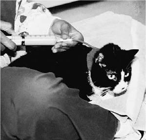
FIGURE 11-4 Esophageal and gastric assisted tube feeding are two vital tools for enhancing quality and length of life in dogs and cats with tumors of the gastrointestinal system. Fluid therapy, medical therapy, and nutritional support can all be achieved through these tubes.They are easy to place and maintain.This cat is being fed via an esophagostomy tube. (Photo courtesy Dr. K.L. Mitchener.)
Because lack of success has often been experienced with other treatment modalities, trials with chemotherapeutic agents have been attempted. Mitoxantrone at doses up to 6.5 mg/m2 intravenously every 3 weeks caused 1 complete remission and 3 partial remissions for 21 to 60 days in 32 cats. In another study, 7 cats received mito- xantrone (5 mg/m2 intravenously every 3 weeks) during and following megavoltage radiation therapy. Radiation was delivered as a “shrinking field,” whereby the tumor and mandibular lymph node received 39.6 to 46.2 Gy, the mandible received 49.5 to 51 Gy, and the gross tumor received 59.4 to 61.2 Gy in daily 3-Gy fractions. The median survival for these cats was 180 days. Thirty percent of the cats were alive 1 year after radiation therapy. Complete remission has been reported in 2 cats with squamous cell carcinoma after treatment with carboplatin. Oral piroxicam therapy (0.14 mg/lb orally every other day) may be of benefit for clinical symptoms and as an antineoplastic agent.
The best therapy for oral squamous cell carcinoma in cats has not been determined. A combination of surgery, radiation therapy, and chemotherapy probably offers the best chance of success. Preoperative radiation and chemotherapy may cause tumor shrinkage and allow surgical margins to be more easily attained.