Parasitic Diseases
Many external parasites, including lice, ticks, and mange mites, infest goats. Each is discussed separately, but there is an extensive overlap in the realm of therapy. For the convenience of the reader, some of the chemicals effective against external parasites are listed in Table 2.1 (Scott 1988; Bowman 2014).
Lice (Pediculosis)
All species of goat lice complete their life cycle on the host and are quite host specific. Eggs (nits) are attached to hairs and hatch in 5-18 days. The nymphal stages look like tiny adults; young lice mature 14-21 days after hatching. Bovicola (Damalinia) ovis, the sheep louse, can become established on goats, and thus goats may be a source for reinfesting sheep during louse control programs (Hallam 1985).
Clinical Signs and Diagnosis
Bloodsucking lice (order Anoplura) have relatively narrow heads with piercing mouth parts. Two species have been reported from goats in the United States: Linognathus stenopsis (females 2.75 mm long, males 2.2 mm) and
Table 2.1 Some chemicals used for control of external parasites of goats.
| Chemical | Concentration and form |
| Amitraz (L) | 0.025-0.05% spray |
| Coumaphos (L) | 0.25% spray, 0.5% dust |
| Crotoxyphos (L) | 0.25-1% spray, 2% dust |
| Dichlorvos (L) | 0.5-1% spray |
| Eprinomectin (L) | 0.5-1 mg/kg pour-on |
| Fenvalerate | 0.05% dip |
| Fipronil | 0.29% spray (not labeled for food animals) |
| Ivermectin | 20-40 mg/100 kg subcutaneous injection |
| Lime sulfur (L) | 2-5% dip (availability limited in USA) |
| Lindane | 0.06% spray, 0.03% dip |
| Malathion | 0.5% spray, 5% dust |
| Methoxychlor | 0.5-1% spray/dip, 5% dust |
| Neem seed extract | 650 ppm aqueous solution |
| Permethrin (L) | 0.05% spray, 0.5% spot treatment |
| Phosmet | 0.15-0.25% spray/dip |
| Trichlorfon | 0.2% spray/dip |
Note that many of these products are not licensed for goats in most countries.
Chemicals designated by (L) are generally appropriate for dairy animals. Product labels should be read for instructions and safety precautions.Source: Based on Scott 1988; Bowman 2014.
Linognathus africanus (distinguished by bulging posterolateral margins of the head; females 2.15 mm, males 1.65 mm). They are bluish-gray and infest both Angoras and the other breeds of goats (Price and Graham 1997). Blood loss and secondary bacterial skin infections may occur in addition to pruritus. Heavy infestations may kill kids.
Biting lice (order Mallophaga) have broader chewing mouth parts. They are pale, small (1-2 mm), and more difficult to see. They often (but not always) provoke rubbing and scratching and can also bite through the hairs, resulting in alopecia and moderate to severe damage to Angora fleeces, depending on the louse numbers. The yellowish, hairy Bovicola (Damalinia) (Holakartikos) crassipes and the red louse Bovicola (Damalinia) limbatus are common on Angora goats, while Bovicola (Damalinia) caprae is the slightly smaller biting louse usually found on meat or dairy goats in the United States. The same goat can be infested with more than one louse species at a time (Sebei et al. 2004). Certain goats appear to have a natural resistance to biting lice, as documented during efforts to cause experimental infestations (Merrall and Brassington 1988).
Diagnosis is by physical examination and demonstration of lice or nits directly or on material collected by plucking or combing. A flea comb works well to harvest material that can be placed in a ziplock bag for later examination with a dissecting microscope or hand lens. In one study, biting lice were concentrated in the withers area (where the goat had more difficulty grooming itself), while sucking lice were more prevalent over the brisket and shoulders (Merrall and Brassington 1988). Because lice are sensitive to elevated temperatures, their location on the body may vary with air temperature and exposure to sunlight.
Populations are generally higher in winter than in summer.Therapy
Numerous insecticides are effective against lice (Moore et al. 1959; Bowman 2014), but nits are not killed. Resistance also may develop, and has been documented for permethrins (Levot 2000), but in some instances may be the result of product failure to reach the lice (Bates et al. 2000). Label directions should be followed to avoid contamination of milk and meat. Because nits are not killed by the initial therapy, treatment should be repeated at 10-14-day intervals if possible to remove young lice before they mature. Treating before kidding helps to prevent the normally rapid transfer of lice to newborn kids. In goat breeds that are shorn, and even in dairy breeds, the best results are obtained by treatment after removal of the fleece and attached nits. All animals in the herd should be treated simultaneously.
Products used include crotoxyphos (1% in water spray or 3% dust), coumaphos (0.25% in water spray or 0.5% dust), dichlorvos, and fenvalerate. Pour-ons are more convenient than sprays or dips, and dusts are preferred to dips in cold weather. Coumaphos (Konar and Ivie 1988) and fenvalerate pour-ons may cause milk contamination. Newer permethrins appear to be both safe and effective, with no milk or meat withdrawal requirements. In India, flumethrin pour-on at 1 mg/kg provided complete louse control on goats for more than six weeks (Garg et al. 1998). Eprinomectin as a pour-on would be safe for lactating goats, but is not approved in the United States. Ivermectin, also unapproved, at 20 mg/100 kg subcutaneously, is efficacious against sucking but not biting lice; it should not be used in lactating dairy goats and this dose is likely to select for resistant gastrointestinal parasites (see Chapter 10). Neem, a botanical extract of the Azadirachta indica tree, is available commercially in some countries and has been found to be helpful for controlling lice on Angora goats (Habluetzel et al.
2007). Likewise, insect growth regulators such as diflubenzuron reduce louse numbers long term on Angoras (Fourie et al. 1995). For pets and young kids, rotenone, flea powders labeled for cats, and even flea collars may be more convenient than commercial livestock preparations. Note that extralabel use of pesticides is not sanctioned in the United States and agricultural products containing rotenone are being withdrawn from the market.Insect hormones have been used experimentally to control lice (Price and Graham 1997). Three spray treatments of 0.1% synthetic juvenile hormone at two-week intervals controlled biting lice for four months (Chamberlain and Hopkins 1971).
Fleas
Dog and cat fleas (Ctenocephalides spp.) sometimes infest goats in tropical regions (Obasaju and Otesile 1980; Opasina 1983)and in the United States (Konnersman 2005b). In Greece, human fleas (Pulex irritans) cause long-term infestations of dairy goats (Christodoulopoulos and Theodoropoulos 2003; Christodoulopoulos et al. 2006). The wingless, laterally compressed, 2-4 mm long adult insects suck blood and cause local irritation. Fleas frequently leave their hosts. Eggs are laid on the goat or on the ground. Clinical signs include restlessness, rubbing and chewing, excoriations, alopecia, non-follicular papules, and crusts. The fleas are easiest to find when the goat is restrained on its back. Anemia and weight loss may occur in young or debilitated animals (Fagbemi 1982) and the infestation may be fatal if not addressed. Peripheral eosinophilia and infiltration of the skin by eosinophils suggest that an allergy to flea saliva may be involved in severe cases (Yeruham et al. 1997). Treatments suggested for lice are also appropriate for fleas, but the environment and all mammalian hosts must be treated.
The sand flea, Tunga penetrans and other Tunga species, is a very small flea, initially 1 mm long, found in Central and South America, sub-Saharan Africa, and India. The larvae live in sand and the adult females burrow into the skin of humans and many other mammals, where they suck blood and breathe, defecate, copulate, and expel eggs through a small orifice in the skin.
Although goats are rarely infested, lameness and localized pain and necrosis have occurred where they penetrated the hoof wall and the skin at the coronary band of kids (Mutebi et al. 2016). Additional goat cases have been reported in South America (Pampiglione et al. 2009).Keds
Melophagus ovinus is a wingless bloodsucking fly (six legs) that infests both sheep and goats. The parasite is 6-7 mm long and easily seen, resembling a tick. The entire life cycle is completed on the host in five weeks or longer. The parasite causes skin irritation and blood loss, but damage to hides may have greater economic significance. Shearing removes many keds. The insecticides used for louse control are also effective against keds. Ivermectin is highly efficacious (Shoorijeh et al. 2007).
Biting Flies, Gnats, Mosquitoes, and FireAnts
Black flies (Simuliidae) may attack goats in swarms in the spring in areas of running water. Insecticides work poorly for repelling these pests, and stabling during the day may provide the most relief (Gnad and Mock 2001). Stable flies (Stomoxys calcitrans) and horse flies and deer flies (Tabanidae) also inflict painful bites. Residual topical permethrin products will provide some relief. Culicoides gnats are additional bloodsucking pests that swarm in the afternoon and evening. They are reported to cause significant skin lesions on small ruminants (Gnad and Mock 2001), but these lesions are not well described. Individual animals may become hypersensitized to the saliva of the gnats. Culicoides gnats are important vectors for bluetongue virus and bunyaviruses. Mosquitoes take blood meals from goats, as from other mammals. Control efforts are usually directed at eliminating stagnant water.
Fire ants, especially the red imported fire ant (Solenopsis invicta), can inflict painful bites on goats and are especially hazardous for neonates and weak animals. They cause scτiL∣ s problems for people and livestock in many warmer parts of the world, including the southeastern United States, Australia, and South America.
Areas lacking long hair (ears, eyelids and eyes, muzzle, perineum, ventral abdomen) are most commonly stung and the necrotizing toxin will produce a papule that develops into a pustule. Remove the animal from the vicinity of the ant mounds and remove any ants from the goat. Ointments containing corticosteroids may provide some relief from the inflammation caused by the stings (Drees 2002).The diagnosis of insect hypersensitivity is usually presumptive in an animal that develops seasonal pruritus and self-excoriation (Figure 2.9). If insect repellants and stabling do not control the signs, injectable dexamethasone can be used in non-pregnant goats.
Cutaneous Myiasis
The New World screwworms Cochliomyia (previously Callitroga) hominivorax in North, Central, and South America are the maggots of flies that must deposit their eggs in wounds or at body orifices rather than in dead carcasses. The species was eradicated from the United States in 1966 by the use of sterile males. The same technique was used after this screwworm was accidentally introduced into Northern Africa in 1988 (Lindquist et al. 1992). The parasite is reportable in the United States and briefly entered Texas from Mexico in 1982; it also arrived on assorted imported animals and people in later years (Alexander 2006). One case involved a larva found on an Angora goat in Texas in 1998, following a hurricane (AVMA 1999). High mortality was observed in Florida Key deer in 2016, but livestock were
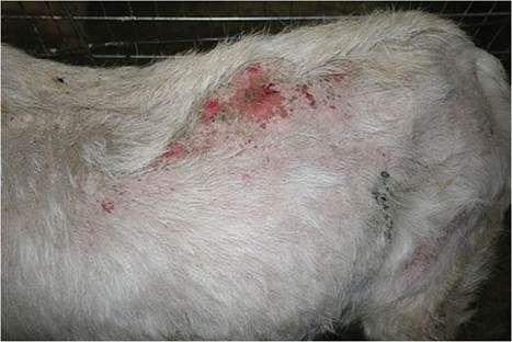
Figure 2.9 Apparent recurrent insect hypersensitivity in a buck that showed severe pruritus and self- excoriation in the early spring in NewYork. Source: Courtesy of Dr. M.C. Smith.
not affected during that outbreak, which was contained by the use of sterile males (Hennessey et al. 2019). Shearing, dehorning, castration and ear-marking wounds, and tick bites are common targets, as are the mouth and navel of newborns. As a complication to the presence of maggots in foul-smelling, pruritic lesions, toxemia or septicemia may kill the goat. Wounds should be debrided and treated with a topical insecticide such as permethrin or malathion or cou- maphos in ointment form. Fly repellents prevent repeat attacks, and antibiotics are indicated if the animal is systemically ill. Injectable avermectins have prevented establishment of screwworm larvae in wounds of calves (Alexander 2006) and oral avermectins were fed to Key deer during the recent (2016-2017) outbreak in Florida (Hennessey et al. 2019). Dicyclanil, an insect growth regulator, shows promise for long-term prophylaxis of this and other forms of myiasis (Sotiraki et al. 2005). Milk withdrawal intervals for parasiticides must be observed in dairy animals, and possible adverse effects on dung beetles should also be considered (Wardhaugh 2005).
Chrysomya bezziana is known as the Old World screwworm fly and has been documented to cause myiasis in goats on the Arabian peninsula (Spradbery et al. 1992; Abo-Shehada 2005; Hall et al. 2009). Although this fly has not yet been transferred to the Western Hemisphere, modern transportation certainly makes translocation possible.
Wohlfahrtia magnifica is an important cause of primary myiasis in Asia (Iran to Mongolia) and the Mediterranean area. Infestation of goats increases with age, and the body regions most commonly struck are the genitalia and the extremities (Ruiz-Martinez et al. 1991; Sotiraki and Hall 2012). Clinical signs include inflammation, pruritus, apathy, and weight loss (Ruiz-Martinez et al. 1987).
A variety of blowfly larvae (calliphorine myiasis) can be found in the same wounds that attract screwworms and is differentiated by close examination of larval anatomy. Bacterial activity (fleece rot, fecal contamination of skin, urine scald) also can make intact skin attractive to these flies. Foul-smelling ulcers are filled with maggots. Cleansing of the wound with a mild solution of pine oil is helpful, because most of the maggots will come out of their holes and drop to the ground. Insecticides, fly repellents, and antibiotics are applied as for screwworms.
Warbles
Przhevalskiana (Hypoderma) silenus is a warble fly that naturally infests goats in Mediterranean countries and Asia. Hypoderma aeratum is reported to parasitize goats in Cyprus, Crete, and Turkey, while Heterocerus crossi infests goats in the dry, hilly regions of India (Soulsby 1982). Recent authors have suggested that these are all one species (Otranto and Traversa 2004). Hypoderma bovis and Hypoderma lineatum (warble flies of cattle) have not been documented to infest goats (Colwell and Otranto 2006).
Pathogenesis and Clinical Signs
Adult Przhevalskiana silenus flies lay eggs on the hairs of the legs and chest in spring. It is believed that the larvae migrate by a direct subcutaneous route to the back rather than passing through the esophagus or spinal cord (Otranto and Puccini 2000). First-stage larvae appear under the cutaneous trunci muscle, which becomes necrotic and infiltrated by neutrophils. The larvae penetrate the overlying muscle and skin and molt into the second instar. Larval debris and cellular infiltrates accumulate, and a wall of granulation tissue forms around each larva (Cheema 1977), which is typically 10-12 mm long. Third-stage larvae drop to the ground to pupate. As many as 150 larvae have been found in a single goat (Prein 1938). In a slaughterhouse survey in Turkey, 53% of 1049 goats examined were infested with P. silenus. Larval numbers ranged from 1 to 52 per infested animal, with an average of 7 (Goksu 1975). In another slaughterhouse survey from Iran, the prevalence of warbles was highest, up to 28%, in goats 6-24 months of age, with none found in goats less than 6 months old (Oryan et al. 2009). Sometimes the hide has the appearance of a sieve. The holes in the hides cause tremendous losses in the fine leather industry in Iran, where 93% of goats in a slaughterhouse study were infested (Rahbari and Ghasemi 1997). Weight loss and poor milk production can also result from heavy infestations (Yadav et al. 2012).
Therapy
Ivermectin, at dosages of 5-20 mg/100 kg, has been effective in killing all instars of the warbles (Tassi et al. 1987; Yadav et al. 2006). Even microdoses of ivermectin (0.5 mg/100 kg as injectable, 1 mg/100 kg as pour-on) are highly efficacious and result in minimal milk contamination (Giangaspero et al. 2003). Modern systemic insecticides are often not available in countries where this parasite abounds. Shepherds have been taught to remove warbles during the winter months.
Ticks
Ticks are important bloodsucking ectoparasites throughout the world.
Etiology
A discussion of the life cycles and identification of the many species reported to infest goats is beyond the scope of this book; readers should refer to standard parasitology texts (Bowman 2014). Some important species are listed in Table 2.2 and species occurring in the United States have been reviewed by Gnad and Mock (2001). Soft ticks belong to the family Argasidae and feed repeatedly on their hosts. Hard ticks, family Ixodidae, have a shield on the dorsal surface and feed only once during each stage. Larval ticks have six legs, while nymphal and adult ticks have eight legs. Ticks are referred to as one-host, two-host, or three-host, according to whether they drop off the host to molt between larval and nymph stages and between nymphal and adult stages. Ticks are not especially host specific (Scott 1988).
Clinical Signs and Diagnosis
The attachment site varies with the tick species (Baker and Ducasse 1968), and may show papules, pustules, or wheals initially, with crusts and ulcers forming secondarily. Demonstration of the tick will make the diagnosis, but skin biopsy to demonstrate embedded tick mouth parts may be necessary in persistent, nodular lesions. Secondary bacterial infections and myiasis are possible. Ticks such as Amblyomma hebraeum and Rhipicephalus glabroscutatum attach to feet, especially between the claws, and may predispose to foot abscesses (MacIvor and Horak 1987). Damage to hides is important. Other important consequences are blood loss and the Irans- mission of very serious diseases such as anaplasmosis, babesiosis, heartwater, Q fever, theileriasis, and tick- borne fever. Tick paralysis may also occur. Pituitary abscess has been associated with attachment of ticks beneath backswept horns of Boer goats in South Africa (Bath et al. 2005). These conditions are discussed else - where in this book.
Table 2.2 Some ticks of importance to goats.
| Tick | Distribution | Importance |
| Argasidae: soft ticks | ||
| Otobius megnini | North and South America, southern Africa, India | Causes irritation and blood loss in ears |
| Ornithodorus spp. | United States, Asia | Transmits Q fever, Theileria, and Anaplasma |
| Ixodidae: hard ticks | ||
| Ixodes ricinus | Europe | Transmits tick-borne fever, louping ill, tick paralysis |
| Ixodes pilosus | South Africa | Does not cause paralysis |
| Ixodes rubicundus | South Africa | Tick paralysis |
| Boophilus decoloratus | Ethiopia | Transmits Borrelia, Theileria |
| Boophilus microplus | Tropics | Tropical cattle tick |
| Rhipicephalus appendiculatus | Africa | Attaches in ears and beneath tail; transmits Nairobi sheep disease |
| Rhipicephalus bursa | Africa and southern Europe | Transmits Theileria ovis, Babesia ovis |
| India | Transmits tick-borne fever | |
| Haemaphysalis punctata | Europe, North Africa | Transmits Babesia motasi, Theileria |
| Amblyomma hebraeum | Africa | Transmits heartwater |
| Amblyomma variegatum | Africa and Caribbean | Transmits heartwater |
| Amblyomma cajennense | North and South America | Tick paralysis (Brazil) |
| Dermacentor spp. | Worldwide | Sometimes causes tick paralysis |
Therapy
Simple extraction of the tick is recommended when ticks and goats are few. A device with a slit that is placed around the mouthparts and then rotated works better than tweezers and direct traction for manual removal (Zenner et al. 2006). A large variety of sprays, dips, and pour-ons (see Table 2.1) will reduce the population of ticks and provide temporary protection. The use of insecticides can be reduced by local spraying or hand-dressing of the body area (ear canal, perineum) where ticks are located (Baker and Ducasse 1968; Schwalback et al. 2003). Systemic ivermectin may also prevent complete engorgement (Wall and Shearer 2001). Residues in meat, milk, and the environment are a concern, as is the possible development of resistance of ticks to insecticides (Stampa 1964) and of gastrointestinal strongyles to ivermectin.
Control
Total eradication of a tick species is usually very difficult. A reduction in tick numbers is achieved by several applications of insecticide at two- to three-week intervals. Two- and three-host ticks require treatments throughout the tick season, while one-host ticks are more likely to be on the host and therefore killed with one or two treatments. Amitraz has been used as a pasture spray to decrease tick burdens (Harrison and Palmer 1981). In certain circumstances, burning the pasture or cultivating the land may aid in tick control.
Sarcoptic Mange
The mite that causes sarcoptic mange (scabies) in goats has been referred to as either Sarcoptes rupicaprae or a goatspecific strain of Sarcoptes scabiei. It tunnels through the epidermis and feeds on tissue fluids (Scott 1988). Experimental infection of desert sheep with a goat strain of Sarcoptes scabiei has been documented (Ibrahim and Abu- Samra 1987) and probably occurs naturally when the two species are raised together. Molecular analyses suggest that all sarcoptic mites belong to a single, heterogeneous species (Zahler et al. 1999). Human infection can occur from handling affected goats (Menzano et al. 2007).
Clinical Signs
The condition is reported to begin with the appearance of small pruritic nodules, especially on the head, several weeks after contact with another infested goat or chamois (Menzano et al. 2007). The skin disease seems to be selflimiting in some goats, while others develop extensive severe dermatitis around the eyes and ears and on the neck and thorax, inner thighs, udder, and scrotum. Hyperkeratosis and alopecia are accompanied by self-excoriation and restlessness. The thickened skin is wrinkled and fissured (Abu-Samra et al. 1981; Kambarage 1992) and may harbor a secondary bacterial infection. Severely affected goats lose weight and have prominent peripheral lymph nodes. Some deaths occur (Zamri-Saad et al. 1990; Menzano et al. 2007; Giadinis et al. 2011). The value of the hide is markedly decreased.
Diagnosis
Confirmation of the diagnosis usually requires deep scrapings at the margin of lesions; the mites are identified by the presence of long, unjointed pedicels on the pretarsi. Even if no mites are seen in multiple scrapings, a biopsy report of eosinophilic dermatitis and tunnels should indicate a presumptive diagnosis of sarcoptic mange (Deorani and Chaudhuri 1965; Scott 1988). In some chronic cases, diagnosis is made by response to therapy. Local regulations may require reporting a positive diagnosis to government authorities.
Therapy
Treatment of lactating animals can be accomplished with repeated (perhaps 5-10) applications of lime sulfur solution every 5-7 days. Two treatments of 0.05% amitraz 7-10 days apart are recommended for controlling scabies in dairy cattle in the United States, and should thus be safe for dairy goats. In non-lactating animals, many different parasiticides have been applied with some success, but relapses are common when treatment is discontinued (Jackson et al. 1983). Sprays often fail to adequately penetrate the long and thick hair coat of unshorn animals. Some animals may need antibiotics for secondary bacterial infections.
More recently, ivermectin (subcutaneously) has simplified treatment, especially because the inconvenience and stress of bathing a disgruntled goat in cold weather are avoided. A cleansing shampoo is recommended to remove crusts, even if ivermectin is given. However, excellent response, including disappearance of the crusts, was achieved in Saudi Arabia when goats were treated with ivermectin (1 mL of a 1% solution per adult of unspecified weight) twice, one week apart. Live mites were recovered from treated animals for at least two weeks, but all goats yielded negative skin scrapings three weeks after the second treatment (Wasfi and Hashim 1986). In another instance, three doses two weeks apart of subcutaneous ivermectin at 0.2 or 0.4 mg/kg were effective (Zamri-Saad et al. 1990). Three treatments 14 days apart with topical moxidectin at 0.5 mg/kg were successful in clearing a severely infected Italian goat herd of the parasite; a higher dose, more appropriate for goats, might have been more rapidly effective (Menzano et al. 2007). Injectable moxidec- tin at 0.2 mg/kg has also been used successfully in chronically infected goats in Greece (Giadinis et al. 2011). For the lactating animal, a single topical application of eprinomectin (Eprinex* pour-on, Merial, Duluth, GA, USA) at 1 mg/kg (twice the cattle dose) might be preferable, because milk residues remain below the limit established for dairy cows (Dupuy et al. 2001), but a withdrawal should still be observed when this drug is given off label.
Chorioptic Mange
Some authors believe that the chorioptic mange mite of goats belongs to the species Chorioptes bovis, while others consider it to be host specific and designate it as Chorioptes caprae (Scott 1988).
Epidemiology
The mite lives on the surface of the skin and feeds on epidermal debris. It may exist on non-clinical carrier goats and can also survive in the environment for as long as 10 weeks (Liebisch et al. 1985). This accounts for the sudden occurrence of mange lesions in closed herds. Mite populations are affected by the environment, with the highest mite numbers and most severe clinical signs in cold weather. In a New Zealand study of feral goats, the prevalence of chorioptic mites was 100% in the winter, although marked lesions were found on the legs of only 5 of 368 goats overall (Heath et al. 1983).
Clinical Signs and Diagnosis
The lesions (non-follicular papules, crusts, alopecia, erythema, and ulceration) are most commonly or initially found on the lower limbs, with occasional spread to udder, scrotum, and perineal region (Scott 2018). Pruritus, as demonstrated by stamping or chewing at the limb lesions, may be obvious, or the alopecia and crusts may appear to be benign (Figure 2.10). Only rarely is the dermatitis caused by Chorioptes generalized to the entire body (Dorny et al. 1994).
Diagnostic samples can be obtained with a flea comb or by skin scrapings; adding rotenone to the mineral oil used for collecting scrapings will prevent the escape of the mites on the way to the microscope (Scott 1988). The mites have short pedicels on their pretarsi. Zinc deficiency and bacterial skin infections are important differential diagnoses that may be present concurrently with mange. If no mites are initially visualized on the surface of scrapings or crusts harvested from the affected skin, the sample can be boiled gently for 10-15 minutes in a 5% potassium hydroxide (KOH) solution to clear it. The mixture is then centrifuged and examined for mites after decanting the solution, or even resuspended in a sugar flotation solution to increase yield.
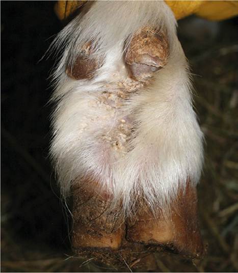
Figure 2.10 Mild crusting and scaling on the pastern of a goat with chorioptic mange. Source: Courtesy of Dr. M.C. Smith.
Therapy
Treatment of chorioptic mange is sometimes very easy, but hypersensitivity of certain goats to the mites can lead to treatment failure and frustration. All in- contact goats should be treated simultaneously to eliminate the carrier state and the premises disinfected. Lime sulfur (four total body sprays or dips in 2% solution, done weekly) is safe for lactating dairy goats. Permethrin can be applied sev - eral times at weekly or biweekly intervals and also sprayed on the pens after removal of bedding. Crotoxyphos (0.25%), coumaphos (0.25%), trichlorfon (0.2%), amitraz (0.05%), and lindane (0.03%) must be applied at least twice at 10-14-day intervals. In one study, a single dip with fenvalerate (0.05%) killed all mites on Angora goats (Wright et al. 1988). Injectable ivermectin can be expected to kill many but not all of the mites, because of their superficial location. Topical application of avermectins such as ivermectin, eprinomectin, or moxidectin is effec - tive against chorioptic mites in other species and is com - monly used to treat goats, but will select for resistant gastrointestinal parasites (see Chapter 10). Fipronil (Frontline* Spray, Merial) has also been advocated for goats (Konnersman 2005b). National regulations concerning milk and meat residues should be respected. Using shampoo to remove crusts and shearing of mohair will improve the efficacy of externally applied acaricides. Systemic antibiotics for secondary bacterial infections and judicious use of glucocorticosteroids in apparently allergic goats are indicated in selected cases.
Psoroptic Mange
The classification of mites of the genus Psoroptes is currently unsettled (Scott 1988; Zahler et al. 1998; Bates 1999). Some propose that Psoroptes recovered from all domestic hosts are conspecific (Pegler et al. 2005). The proposed role of ear mites in the transmission of mycoplasma infections is discussed in Chapter 9.
Etiology and Epidemiology
The most commonly reported ear mite of goats is the cosmopolitan Psoroptes cuniculi. Prevalence in necropsy studies appears to be high (21 of 24 and 8 of 18; Williams and Williams 1978; Cook 1981). The mite can be found in the external ear canal of kids as young as 10 days old, with most kids infested by the third week (Williams and Williams 1978). In feral goats in New Zealand, the infestation was often unilateral and was more common in winter and in older goats (Heath et al. 1983). When it causes body mange (in parts of the world where sheep are affected with psoroptic mange), the parasite is sometimes referred to as Psoroptes caprae. The mite has long, segmented pedicels and is often visible to the naked eye. It does not burrow, but feeds on tissue fluids. Prolonged survival times (as long as 12 weeks) in the environment have been reported (Liebisch et al. 1985).
Clinical Signs and Diagnosis
Clinical signs of ear mite infestation include head shaking (Dorny et al. 1994) and scratching, sometimes with alopecia of the part of the ear repeatedly brushed by a hind foot. Occasional goats have flaky or scabby lesions or laminated crusts on the external ear or even on the poll, back, and pasterns (Littlejohn 1968; Munro and Munro 1980; Heath et al. 1983; Lofstedt et al. 1994), but most have no externally visible lesions. Otoscopic examination (tranquiliza- tion advised for adult goats) easily demonstrates the presence of the mite. A plug of yellowish wax is commonly found in the ear canal (Munro and Munro 1980; Cook 1981; Heath et al. 1983), and extracting some of this material with a cotton swab for examination is an alternative means of diagnosis. Palpation of the base of the external ear may elicit a crackling sound associated with exudate in the canal (Nooruddin and Mondal 1996). Rarely, otitis externa progresses to otitis media and interna with head tilt.
Body mange caused by Psoroptes resembles sarcoptic mange, but is accompanied by less scab formation (Wasfi and Hashim 1986). Before sheep scab due to Psoroptes ovis was eradicated from Texas, Angora goats were noted to have a much more severe form of Psoroptes cuniculi infestation than was seen on dairy goats, as evidenced by serious damage to skin and hair (Graham and Hourrigan 1977). Skin scrapings should be collected from the margin of lesions. If the scrapings are collected into a ziplock bag or petri dish, the sample can be warmed to stimulate movement of the mites during later close examination. Reporting to regulatory authorities may be required.
Therapy
Treatment of ear mange is often forgone or limited to removal of any bells from goats showing clinical evidence of pruritus (to avoid continual disturbance of the owner). External lesions may be swabbed with an acaricide. Canine ear mite preparations may be used on goats with mites confined to the ear canal, but removal of crusts and debris is necessary and recurrence is likely (Munro and Munro 1980). The life cycle probably takes about three weeks, and several weekly treatments are advised (Littlejohn 1968). In non-lactating pet goats, a rational therapy is two treatments with injectable or oral ivermectin one to two weeks apart (20 mg/100 kg; Lofstedt et al. 1994). Instillation of several drops of an ivermectin solution into each ear canal is also effective (Konnersman 2005b).
Body mange can be treated with routine acaricides (including amitraz; Harrison and Palmer 1981) as dips or sprays. Ivermectin (two injections one week apart) also has been found effective in treating psoroptic mange in goats (Wasfi and Hashim 1986).
Raillietia
A separate genus of ear mites of goats is Raillietia (Cook 1981; Lavoipierre and Larsen 1981). The feral goats that harbored only these mites showed no evidence of associated clinical disease, although ear irritation might be expected to occur. Raillietia mites tend to be larger than Psoroptes, and their longer legs originate from the anterior half of the body. The possible involvement of ear mites in the transmission of mycoplasma infections is discussed in Chapter 9.
Demodectic Mange (Demodicosis)
Demodex caprae is a cigar-shaped mite that commonly inhabits the hair follicles and sebaceous glands of goats. It has a worldwide distribution. As in other hosts, demodec- tic mites have been found in the eyelids of goats, with no visible lesions anywhere on the body (Himonas et al. 1975).
Epidemiology
Confinement housing and crowding seem to favor the development of demodectic mange. The mites appear to survive only a few hours away from the goats, but infection via contact with contaminated feeders might occur. In fact, natural spread from one adult goat to another has not been documented. The appearance of nodules in certain goats in a herd but not in others may reflect the immune competency of the animal (Das and Misra 1972). This could be related to genetic factors, nutrition (e.g., selenium or protein deficiency), or stress (e.g., high production). It seems likely that goats are first infested as young kids, but that lesions do not become detectable until many months later (Williams and Williams 1982).
Clinical Signs and Diagnosis
Portions of the body exposed to friction, such as the face, neck, shoulders, and sides, are commonly involved, whereas the ventral abdomen and udder remain free of nodules. Where animals with skin lesions have been followed, it has been observed that nodules are first detectable at 10-15 months of age (Euzeby et al. 1976). Initially the lesions are very small and only located by careful palpation (Smith 1961). However, defects may be readily detectable in hides at this stage or even earlier (Rohrer 1935). By 18-20 months of age the nodules have enlarged to lentil or pea size, but because they remain painless they may not be noticed unless the goat is clipped (as for a show). Occasional animals demonstrate mild pruritus (Durant 1944), although this is a relatively rare finding.
Each nodule corresponds to a distended pilosebaceous follicle. Firm digital pressure will force out a ribbon of yellowish white paste from a central pore. Microscopic examination of this caseous material reveals numerous eggs, larvae, and mature mites (Figure 2.11). The diagnosis is thereby confirmed. The disease process peaks at about 3 years of age. Nodules may reach a diameter of 0.5-1.25 cm.
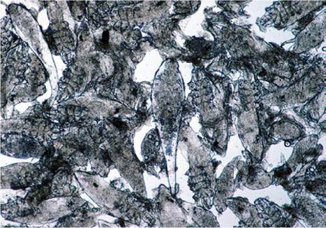
Figure 2.11 Cigar- shaped demodectic mange mites in a smear of exudate expressed from a skin nodule. Source: Courtesy of Dr. M.C. Smith.
At this time, the severely affected goat may be apathetic and unthrifty, and may lose weight or milk poorly. It is not clear if these signs are because of mange infestation or underlying (nutritional) problems. The nodules become smaller, firmer, and less numerous in older animals.
Therapy
Various treatments have been proposed. When only a few nodules are present, squeezing or incising each one to permit removal of its contents will result in a cure of the hair follicle thus treated. Iodine or other disinfectant is applied when the nodule is expressed to kill residual mites. Some goats carry several hundred discrete lesions. Others have a generalized dermatitis rather than the typical nodule form of the disease. For them, topical or systemic organophosphates, amitraz (0.025%), or weekly ivermectin or eprinomectin (for 6-12 weeks) may be effective (Thompson and Mackenzie 1982; Strabel et al. 2003). Because the lesions resolve over weeks or months, documentation of a treatment effect is difficult. Nodules may remain long after the mites have died. In one instance, nodules reappeared after amitraz (12 topical treatments) was discontinued (Brugger and Braun 2000). Because secondary bacterial infections are generally absent in undisturbed lesions, antibiotic therapy is not indicated.
Treatment is normally only undertaken in show animals, except when nutritional deficiencies exist. It has been suggested that severely affected animals should not be used for breeding because of a possible hereditary susceptibility to the disease (Scott 1988).
Free-Living Mites
Trombiculid mite adults and nymphs are free-living. The larvae (chiggers, harvest mites) are reddish and six-legged. They attack the pasterns, muzzle, and ventrum of animals pastured in infested fields and woods, or when contaminated forage is fed to housed animals. Clinical signs of irritation and pruritus, papules, edema, exudation, and ulceration may be expected. Skin scrapings to identify the larvae permit differentiation from other conditions such as chorioptic mange, staphylococcal dermatitis, and zinc deficiency. Mites may be absent in chronic cases. The condition is well reported in sheep (Wall and Shearer 2001) and is occasionally recognized in goats (Nooruddin et al. 1987; Faccini 2017). An insecticide dip or spray might give eventual relief.
Exposure to biting grain or forage mites such as Tyroglyphidae spp. may occasionally cause dermatitis in goats (Matthews 2016). Likewise, a pruritic goat housed with poultry, especially in late summer, might be being attacked at night by poultry mites (Dermanyssus gallinae; Matthews 2016).
Rhabditic Dermatitis and Strongyloidiasis
Strongyloides papillosus larvae penetrate unbroken skin, enter capillaries, and travel in the blood to Ihe 1 ungs. I Ieire they ee^it into air passages and travel up the trachea and down the gastrointestinal tract to the intestines. There are no histologic changes in the skin on first exposure, but pustular derImUtiii^ is created as host resistance develops via repeated c^[p'o>ure;>. This is characterized by edema, inflammatory infiltration (neutrophils, eosinophils, lymphocytes, and giant cells), and destruction of the larvae. This condition is best described in sheep (Turner et al. 1960), but has been created experimentally in goats (Pienaar et al. 1999). Affected goats are Teporteed to stamp, dance, and nibble at their feet, especially after rain (Baxendell 1988). Warm, moist habitats for the la.rva.e should be eliminated and the animal treated with an anlhelminlihc such as fenbendazole, levamisole, or ivermectin.
Pelodera (Rhabditis) Strongyloides is a free-living soil nematode that can invade portions of skin in contact with moist ground and decaying organic matter (Scott 1988). Although not reported in goats, it could cause pruritic dermatitis in goats housed under unsanitary conditions. Diagnosis is by demonstration of motile larvae in skin scrapings or sections of larvae in hair follicles in samples taken for skin biopsy. Control is by sanitation, including removal of dirty bedding. The hookworm (Bunostomum) is another nematode that could conceivably produce dermatitis while penetrating the skin. Additional conditions to be considered in the differential diagnosis include contact dermatitis, mange, and trombiculidiasis (chiggers).
Parelaphostrongylosis and Elaphostrongylosis
Parelaphostrongylus tenuis, the meningeal worm of the white-tail deer, commonly causes neurologic disease in goats in North America. Some paretic and non-paretic goats with P. tenuis infection have developed linear, vertically oriented skin lesions on the neck, shoulder, thorax, or flank (Smith 1981; Scott 1988, 2018). Owners report that the goat has excoriated these areas by biting or rubbing, as if responding to intense pruritus. Lesions are usually unilateral and alopecic, crusted, or scarred (Figure 2.12). One lesion may heal and another appear closer to the goat's head. One possible explanation is that migrating parasite larvae irritate dorsal nerve roots supplying individual dermatomes. The diagnosis of P. tenuis-induced dermatosis should be entertained when goats with linear pruritic lesions have been exposed to pastures frequented by deer. Neurologic deficits suggesting spinal cord damage and eosinophilia or increased protein in the cerebrospinal fluid lend additional support to the diagnosis. Treatment and control of this parasite are discussed in Chapter 5.
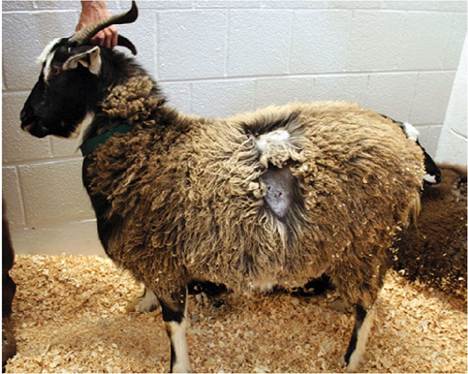
Figure 2.12 Vertically oriented alopecic lesion on the side of a cashmere goat that had been pastured with white -tail deer, suggesting irritation of a dorsal nerve root by Parelaphostrongylus tenuis. Source: Courtesy of Dr. M.C. Smith.
Goats in Scandinavia have been affected with a very similar neurologic and sometimes pruritic disease, in which the migrating parasite is Elaphostrongylus rangiferi and the natural host is the reindeer (Davidson et al. 2020). In one herd pruritus was noted to precede the neurologic signs (Handeland and Sparboe 1991). When goat kids were experimentally infected with E. rangiferi, pruritus was common from 4-10 weeks after infection (Handeland and Skorping 1993). The muscle worm of deer, Elaphostrongylus cervi, has been associated with neurologic disease of goats in Europe, but pruritic skin lesions have not been reported.
A differential diagnosis for this syndrome might be psychogenic self-mutilation (Yeruham and Hadani 2003). Examination of biopsy samples reveals only the skin damage induced by chewing or scratching. However, unless the spinal cord is also examined, it will not be possible to rule out irritation to a nerve root supplying the affected skin.
Filarid Dermatitis
Several filarid worms are known to cause dermatitis in small ruminants. Insect vectors, especially flies, deposit larvae in skin wounds. Papular, alopecic, or crusty lesions develop, and are accompanied by pruritus. In Malaysia, a crusty dermatitis of the feet of goats has been ascribed to Stephanofilaria kaeli (Fadzil et al. 1973). In India, Stephanofilaria assamensis has been recovered from skin sores on goats (Patnaik and Roy 1968). Skin scrapings, and smears of blood oozing from the skin after the scrapings have been made, reveal adult parasites and microfilaria. Biopsy samples may be fixed in formalin or macerated and examined with a dissecting scope. Eosinophils predominate in the inflammatory reaction to the parasites. Trichlorfon has been efficacious for treating affected goats.
Elaeophora schneideri (a subclinical intra-arterial parasite of mule deer and black-tailed deer in the mountains of the western United States) occasionally infests sheep, elk, and moose. Transmission is by tabanid flies (deerflies and horseflies). Adult worms block arteries of the head, and microfilaria lodge in skin capillaries, where they might be demonstrated in a skin biopsy. Erythema, alopecia, ulcers, and crusts appear on the face and poll, and sometimes the abdomen and feet of sheep. Blindness, neurologic signs, and keratoconjunctivitis also may occur. The absence of documented caprine cases may reflect how rarely goats are grazed above 2000 m in the mountain ranges. However, the parasite has been reported in the southeastern United States, Texas, and the Pacific coast states, and goats are listed as occasionally infected (Haigh and Hudson 1993; Klei 2019). One author reports that Elaeophora is highly pathogenic to goats, with more than 10 nematodes resulting in brain damage and associated signs (Anderson 2001).
Besnoitiosis
In Africa and the Middle East, protozoal parasites of the genus Besnoitia cause a widespread dermatitis and alopecia in wild and domestic goats. Some workers believe that the organism infecting goats, termed Besnoitia caprae, is distinct from that infecting cattle, Besnoitia besnoiti (Njenga et al. 1995; Oryan and Azizi 2008; Oryan et al. 2014). Lesions may be especially severe on the scrotum and lower limbs. The skin is thickened to corrugated (hyperkeratosis), sometimes with hyperpigmentation and exudation (Oryan and Sadeghi 1997). Diagnosis is by demonstration in skin biopsy specimens of cysts (oval or spherical, average size 175 ? 290 μm) containing bradyzoites and surrounded by a collagen capsule (Bwangamoi 1967; Cheema and Toofanian 1979). Oryan et al. (2014) report a cyst diameter of 400-800 μm. There is also a correlation with the presence of Besnoitia cysts in the scleral conjunctiva (Bwangamoi et al. 1989). No specific treatment is available. Control measures for goats might include culling of infected animals (Oryan et al. 2014) and avoiding feed contamination with cat feces. Direct contact and biting arthropods possibly transmit the parasite as well (Oryan et al. 2014).