Parasitic Pneumonia
Verminous pneumonia resulting from one or more species of lungworms occurs in most parts of the world. Liver flukes and tapeworm cysts have more restricted ranges.
Dictyocaulus Pneumonia
Etiology and Epidemiology
Although Dictyocaulus filaria is cosmopolitan, in many parts of the world this lungworm is not very common in goats.
This may be caused in part by the tendency to keep young goats off pasture under intensive management systems. In India, sheep and goats graze the same infested pastures, and goats are believed to be more susceptible to the parasite (Dhar and Sharma 1978a).Pathogenesis
Dictyocaulus filaria lungworms have a direct life cycle (Panuska 2006; Bowman 2014). Adults live in bronchi and are approximately 30-80 mm long. First-stage larvae approximately 550 μm long are coughed up and eliminated in the feces. Development to an infective stage takes one to two weeks. These third-stage larvae are long-lived in damp, cool surroundings. After ingestion, there is a one-month prepatent period and three months patency. Signs can occur as early as three weeks after the goats are put on pasture in the spring, but usually occur in autumn. Kids are most severely affected during their first season on pasture; age resistance is via prior exposure (Wilson 1970). The L4 can last the winter in lungs, but freezing kills L3 larvae on pasture, as does a hot, dry summer.
Clinical Signs
The main sign during the prepatent period is polypnea; dyspnea develops later in the course of infection. The worms cause bronchial irritation, resulting in cough (often paroxysmal; Fereig et al. 2018), moderate dyspnea, and loss of condition. Pathologic findings include bronchitis, bronchiolitis, atelectasis, and emphysema. Auscultable crackles and wheezes, fever, and toxemia occur if there is a secondary bacterial infection. Eosinophilia is marked in re-exposed animals.
An acute shock syndrome is seen when a significant reinfestation occurs after immunity has weakened.Diagnosis
The diagnosis of verminous pneumonia is best done by the Baerman technique. Feces are collected fresh from the rectum to avoid confusion with Strongyloides larvae. Fecal pellets are put in a tea strainer or wrapped in gauze and half submerged in a funnel of warm 25 °C (77.7 °F) water; larvae fall to the neck of the funnel. A conical flask may provide a better yield than the traditional funnel system (McKenna 1999). The larvae are harvested after 4-20 hours and compared with photos in a parasitology text. The posterior is blunt in Dictyocaulus, whereas it is slender and pointed in Protostrongylus and has a definite dorsal spur in Muellerius (Bowman 2014). Fecal exams for Dictyocaulus are positive during the patent period of the first infestation only.
Treatment and Control
Anthelmintics effective against Dictyocaulus are routinely used in goats, which probably decreases the prevalence of the disease. Treatments include tetramisole (15mg∕kg; Kadhim et al. 1972) or levamisole (7.5 mg/kg) orally or SC. The drug is eliminated by the respiratory tract. Also reported to be effective are mebendazole (15-20mg/kg), fen- bendazole (5-10 mg/kg), febantel (5 mg/kg), and ivermectin (0.2 mg/kg). Owners should be advised not to sell milk or make it into cheese for at least four days after unapproved drugs are used. The milk withdrawal period for ivermectin should probably last at least 9 days after oral use and 40 days after subcutaneous injection (Baynes et al. 2000). Higher doses of these drugs may be preferable to avoid selection for resistant gastrointestinal parasites; see Chapter 10.
Treatment does not allow establishment of immunity, so goats should not be returned to infected pastures. Frequent pasture rotation is often an impractical means of control because moving the goats every four days would be required. Putting the goats out on pasture early in the season allows progressively increasing exposure and immunity.
New goats should be separated from yearlings, and kids should not be placed on pastures inhabited by yearlings within the last year. The goats are treated after stabling in the fall (a warm, humid barn allows the cycle to continue) to limit pasture contamination next spring. Other suggestions include avoiding overpopulation, draining wet pastures, and replacing ponds with watering devices on concrete slabs. An irradiated Dictyocaulus larvae vaccine has protected goats against experimental challenge (Dhar and Sharma 1978b; Sharma 1994).Protostrongylids
Etiology
Protostrongylid lungworms include at least five genera. Two are well recognized in the United States; Muellerius capillaris is diagnosed more frequently than Protostrongylus rufescens and is also found in surveys of goats around the world. Two more (Cystocaulus ocreatus and Neostrongylus linearis) are recognized in Europe and northern Africa (Genchi et al. 1984; Perreau and Cabaret 1984; Berrag and Urquhart 1996), while Varestrongylus pneumonicus (Bicaulus schulzi) is found in eastern Europe and India.
Epidemiology and Pathogenesis
All types have an indirect life cycle and use a variety of snails and slugs as intermediate hosts. The mollusks remain infective for at least one year. Goats and sheep become infected by eating mollusks or third-stage larvae liberated on foliage by death of the intermediate host.
Protostrongylus rufescens adults live in bronchi and are approximately 16-35 mm long. The first-stage larvae are 370-400 μm long and have a pointed tail (Gerichter 1951). Massive infections are unlikely, and only kids show any clinical involvement.
Muellerius lungworms usually cause clinically insignificant focal lesions in the lung parenchyma. Adults are present 25-38 days after infection (Panuska 2006). They are 12-23 mm long and located in subpleural alveoli, never in the bronchi. First-stage larvae approximately 300-320 μm long (Gerichter 1951) with a distinctive dorsal spur on the tail (Figure 9.8) are passed in the feces if both sexes are present in the same nodule in the lung.
The infestation is carried over from one year to the next in the goat. Fewer adult worms are established following reinfection, but the inflammatory response in the lungs is much more severe (Berrag et al. 1997). Goats do not develop a marked age resistance, and in fact older animals often have heavier infections than those on pasture for only one season. This is in contrast to sheep, which tend to clear the infection.At least 40 species of snails and slugs serve as intermediate hosts for Muellerius. A French study found abundant Helix aspersa in spring and Deroceras reticulatum in autumn to be associated with an increased risk of infection in grazing goats (Cabaret et al. 1986). Almost any goat that has been on pasture in temperate regions of the United States has Muellerius larvae on Baerman examination
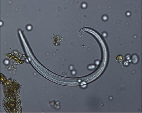
Figure 9.8 Larva of Muellerius capillaris, showing dorsal spur on tail. Source: Courtesy of A. Lucia - Forster.
(see above). In fact, these larvae are also readily found in a routine fecal flotation with centrifugation, without resorting to a Baerman test. In a Norwegian study, larvae were found in the feces of 447 out of 457 goats from seven flocks (Helle 1976).
Cystocaulus ocreatus has a very similar life cycle to Muellerius. It also forms nodules in the lung parenchyma, and uses many of the same mollusks as intermediate hosts. The first-stage larvae are 390-420 μm long. The larvae are identified by a distinctive tail tip with two appendages (Gerichter 1951). The two species may be found simultaneously in the same goat.
Clinical Signs and Necropsy Findings
Muellerius infestations are usually subclinical. However, there are scattered reports of serious, even fatal pneumo - nia resulting from heavy Muellerius infestations. For instance, Lloyd and Soulsby (1978) found 20 of 24 herds infected with Muellerius. Of 169 goats in these herds, 83% were passing larvae, including 100% of goats 3 years of age or older.
Dyspnea and a persistent cough were noticed in four adult goats with higher than average larval counts, suggesting that the parasite may be pathogenic in heavily infested goats. In a Swiss herd of CAE-free Saanen goats, all nine adults displayed coughing that abated only temporarily with anthelmintic treatment. Many Muellerius larvae were present in the feces of all and in several goats first-stage larvae and neutrophils were demonstrated in an increased amount of viscid bronchial secretion by endoscopy (Braun et al. 2000b). Hematology and serum biochemistry test results were unremarkable in these goats. A possible association of Muellerius with decreased bodyweight has been reported in New Zealand (Valero et al. 1992).Lesions (firm tan nodules) are most common in the caudal dorsal lung lobes and are less well localized in goats than in sheep. Sometimes the lesions are greenish in color, presumably due to the presence of eosinophils. A frequently cited report (Nimmo 1979) ascribed a diffuse thickening of alveolar septa with mononuclear cell infiltrates to Muellerius. That paper suggested that diffuse (non-nodular) lesions occurred in cases of reduced host resistance, because eosinophils were more prominent in focal nodular lesions. Because L1 larvae, eggs, and adult worms in “brood nodules” are readily identified in histologic preparations of the lung (Gregory et al. 1985), it must be remembered that other conditions (such as progressive retroviral pneumonia) must be excluded before ascribing a clinically important pneumonia to these parasites. Also worth considering is the possibility that Muellerius lungworms contribute to clinical retroviral infection by attracting many macrophages to the lungs (Ellis et al. 1988a).
Treatment
Recommended treatments for Protostrongylus include lev- amisole and fenbendazole at dosages used for Dictyocaulus.
Levamisole is not effective against Muellerius. When considering drugs and dosages for Muellerius given in the literature, it is not always evident if efficacy was determined by death of the protostrongylids or a temporary infertility of the worms (Cremers 1983).
Two treatments with fenbendazole at 15 mg/kg, three weeks apart, relieved clinical signs and completely eliminated shedding of larvae two weeks later (Kazacos et al. 1981). Extended studies revealed that larval shedding resumed in most goats by seven weeks after 30 mg/kg fenbendazole (Bliss and Greiner 1985). Another study found that shedding of larvae resumed many weeks after treatment with either fen- bendazole or ivermectin; 25% of ivermectin-treated animals resumed shedding from four to more than nine weeks later. The authors assumed that immature Muellerius in the lungs resumed development after death of adult worms (McCraw and Menzies 1986, 1988). Treatment at approximately 35-day intervals has been suggested. Topical eprinomectin at 0.5 mg/kg was shown to prevent larval shedding for six weeks or maybe more (Geurden and Vercruysse 2007). A long-acting injection of moxidectin decreased larval shedding by 99% for at least 77 days (Vadlejch et al. 2016). Netobimin at 20 mg/kg (once orally or divided into two or three doses) failed to completely eliminate shedding or kill all adult Muellerius at day 18 (Cabaret 1991). For other drugs such as mebendazole (20-40 mg/kg) and oxyfendazole (7.5-10 mg/kg) (Cabaret et al. 1984), the dose suggested is typically double that used for digestive strongyles.Control
In Scandinavia, a protocol has been developed to specifically control Muellerius in goats. During the period when the goats are stabled and not lactating, fenbendazole is fed at 2 mg/kg/day for 14 days. Prolonged low-level treatment reduces larval shedding for five to seven months (Helle 1986). This treatment is believed to decrease the prevalence of coughing and increase subsequent milk production and bodyweight gains (K.E. Hammarberg, Hudiksvall, Sweden, personal communication, 1992). The possible teratogenic effects of anthelmintics have not been well studied in goats, and it is prudent to avoid treatment during the first 35 days of pregnancy. Also, such a treatment program should not be undertaken when the goats are on pasture, because it might select for resistant gastrointestinal parasites.
Wet, undrained pastures should be avoided. It may be helpful to prevent grazing in early morning or evening when the herbage is damp and snails are up and about. It is best to treat before pasture season in the spring, to prevent contamination of mollusks. A second treatment in early fall is used in heavily contaminated environments. To date, the nematophagous fungus Duddingtonia fla- grans has not been shown to be effective in decreasing the survival of Muellerius larvae on pasture (Paraud and Chartier 2003).
Eimeria and Other Protozoa
A persistent cough is often associated with severe coccid- ial infection in young kids, though no adequate explanation has been advanced. The connection may be only that kids stressed by the poor nutritional state that accompanies coccidiosis are more prone to bacterial pneumonia. Prophylaxis includes keeping kids out of feeders, separate from adults, and in a dry environment. A systemically absorbed sulfonamide would be reasonable treatment for such kids.
There is a single case report of the presence of cysts and trophozoites of Balantidium coli in the lungs of a goat with interstitial pneumonia and D. filaria larvae (Parodi et al. 1985). Because this ciliated protozoon normally inhabits the large intestine, it might have been carried to the lungs by lungworm larvae.
Besnoitia cysts (diameter 60-280 μm) were reported in the alveolar septa of the lungs of two adult goats from Kenya with purulent necrotizing bacterial pneumonia (Kaliner 1973). It has been proposed that the species of Besnoitia infecting goats, B. caprae, is distinct from that infecting cattle, at least in Kenya (Njenga et al. 1993).
Echinococcosis, Hydatidosis
Echinococcus granulosus hydatid cysts occur in goats in many parts of the world, especially Mediterranean countries, India (Upadhyaya et al. 1983), and Africa. Dogs are the definitive host and herbivores or humans are intermediate hosts. Goats appear to be less susceptible to infection than sheep in a given area, possibly because browsing causes less exposure to dog feces than does grazing (Rausch 1995). Goats have been infected by both sheep and cattle strains of the parasite (Rajabloo et al. 2012). Cysts are most often observed in liver and lungs in goats (Pandey 1971; Perreau and Cabaret 1984), and the condition is discussed in greater detail in Chapter 11. Transplacental infection is possible. The cysts can be as large as a fist, but are typically pea to plum size. The surface of the lung is distorted by focal collapse and emphysema. Older goats have more and larger cysts. They rarely cause specific clinical signs, but can be demonstrated by ultrasound examination (Njoroge et al. 2005). No easily administered treatment has been found effective against the parasite in goats (Thompson and Allsopp 1988). Oxfendazole given orally at 30 mg/kg twice a week for four weeks resulted in the death of 14 of 15 cysts in a group of three goats and two sheep (Njoroge et al. 2005).
Prevention of zoonotic infection requires breaking the cycle between definitive and intermediate hosts (Gemmell 1979). All dogs should be treated (preferably with praziquantel) and feces passed after treatment should be destroyed. Eggs excreted in feces of dogs remain infective for at least one year. Wild dogs must be controlled. Access by dogs to carcasses of ruminants that die or are slaughtered on the farm or at the local slaughterhouse must be prevented. These measures are very difficult to enforce. More recently, vaccination of ruminants using a recombinant oncosphere peptide combined with twice-a-year dog dosing has shown promise (Craig and Larrieu 2006). Lambs and sheep are vaccinated twice at a one-month interval, with a booster 6-12 months later. Although few reports are available for the use of this vaccine in goats, kids have been protected with two doses given a month apart (Heath et al. 2003).
The dog tapeworm Taenia multiceps (Multiceps gaigeri) has also caused cysts within the thoracic and abdominal cavities and in muscles of goats in the Sudan (Hago and Abu-Samra 1980).
Liver Flukes
Both Fasciola hepatica and Fascioloides magna occasionally invade the lung. Their large size and the dark pigment that often accompanies them make the diagnosis at necropsy simple. Accompanying liver disease would be expected to overshadow any clinical signs originating from the lungs.
Schistosomosis
Pulmonary schistosomosis (Schistosoma indicum) has been reported in goats in India (Sharma and Dwivedi 1976a; Dadhich and Sharma 1996). Granulomas occur diffusely throughout the lung where adult parasites have laid eggs in pulmonary vessels. Emaciation and dyspnea may occur. The lesions grossly resemble retroviral interstitial pneumonia. Schistosomosis is discussed in Chapter 8.