Taxonomy of Lacazia loboi and Paracoccidioides brasiliensis var. ceti
Paracoccidioides brasiliensis (Splendore) de Almeida var. ceti Vilela, St. Leger, Bossart, and Mendoza, var. nov.
Holotype B92-932, H&E histopathological slide collected from a US dolphin, deposited at the Michigan State University Herbarium, East Lansing.
Etymology To differentiate this novel uncultivated variety of P. brasiliensis affecting dolphins from the cultivated type causing human systemic paracoccidioidomycosis, the variety ceti (Cetacea) is proposed.
Description Uncultivated fungus causing cutaneous disease in dolphins. In vivo, numerous branching chains of globose to subglobose, yeast-like cells (5-10 μm) present, some connected by slender bridges (2-3 μm). Adjacent older cells detach by increasing cell wall thickness.
Disease nomenclature We propose the name “paracoccidioidomycosis ceti” to differentiate this cutaneous disease of dolphins from human systemic paracoccidioidomycosis (Vilela et al. 2016).
Due to the unculturable nature of L. loboi and P. brasiliensis var. ceti, the taxonomy of these two etiologic agents of cutaneous granulomas has been contentious (see above). Lacazia loboi from humans and P. brasiliensis var. ceti from dolphins share several features: (1) they resist culture (Lacaz et al. 1986; Schaefer et al. 2016); (2) they are restricted to cutaneous granulomas in mammalian hosts (Baruzzi et al. 1973; Bossart et al. 2015); (3) they develop similar yeast-like cells in chains (Haubold et al. 2000; Vilela et al. 2016); and (4) they possess cross-reactive antigens (Mendoza et al. 2008). Using traditional approaches, these two mammalian pathogens were believed to be the same organism. Only recently, with the use of molecular methodologies, their true position in the tree of life was unveiled (Herr et al. 2001; Rotstein et al. 2009; Vilela et al. 2016).
Initially Herr et al. (2001) extracted total L.
loboi DNA from a biopsied tissue collected from a Brazilian man with cutaneous lacaziosis. They found that ITS, chitin synthase 4 (CHS4), and 18S SSU rDNA sequences placed this pathogen as a sister group to P. brasiliensis, the etiologic agent of systemic paracoccidioidomycosis. The authors argued that their phylogenetic data confirmed Lacaz (1996) position on the similarities shared by these two pathogens. The main problem studying the phylogenetics of L. loboi, however, was that the total DNA recovered from biopsied tissues contained DNA from the host and from normal skin and environmental microbiota. Vilela et al. (2005) proposed a molecular model to study this unculturable pathogen using specific primers. They argued that due to their phylogenetic proximity, L. loboi might share common DNA sequences with P. brasiliensis. Using this approach, they were able to amplify the gp43-like gene in L. loboi using well-known DNA sequences of this gene in P. brasiliensis. This approach was later validated when Vilela et al. (2009) amplified the ITS rDNA and chitin synthase 4, ADP-ribosylation factor, and gp43 coding genes. The phylogenetic data showed that indeed L. loboi clustered with strong support in its own genus, confirming previous analysis (Vilela et al. 2009). These analyses showed that P. brasiliensis and P. lutzii shared the same ancestor with L. loboi and P. brasiliensis var. ceti; thus the latter unculturable microbes may possess putative dimorphic capabilities.When Vilela et al. (2009) concluded their studies, L. loboi was still considered the same etiology affecting both humans and dolphins. However, Rotstein et al. (2009) found that the DNA sequences (26S LSrDNA) recovered from an offshore dolphin
(T. truncatus) with cutaneous granulomas displaying yeast-like cells shared 97% identity with P. brasiliensis. Unfortunately, their DNA sequences are not available. Subsequently, two teams in Japan (Minakawa et al. 2016; Ueda et al. 2013) and one in Spain (Esperon et al.
2012) reported that the DNA sequences, using at least two types of sequences (ITS and gp43), placed the etiologic agent of dolphin granulomas within the DNA sequences of the culturable P. brasiliensis isolates causing human systemic paracoccidioidomycosis. These findings came as a surprise since the clinical morphological features of the yeast-like cells in the dolphin-infected tissues look similar to that in human lacaziosis (Haubold et al. 2000; Lacaz et al. 1986). To validate these findings, Vilela et al. (2016) amplified the coding kex gene from six dolphins (T. truncatus) with cutaneous granulomas collected and two additional CHS4 DNA sequences from different dolphins at the Indian River Lagoon, Florida, USA (Fig. 9.1). They confirmed previous studies (Esperon et al. 2012; Minakawa et al. 2016; Rotstein et al. 2009; Ueda et al. 2013) and concluded that an unculturable type of P. brasiliensis, different from the culturable type causing human systemic infections, is the etiologic agent of cutaneous granulomas in dolphins. They named the disease paracoccidioidomycosis ceti, epithet used throughout this chapter.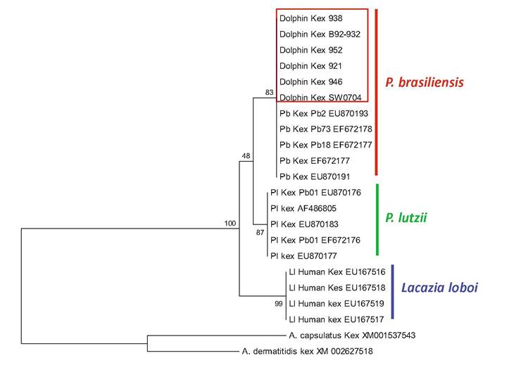
I------------------- 1
5
Fig.9.1 Maximum parsimony tree of the exon partial kex DNA sequences PCR amplified from six US dolphins. The phylogenetic tree depicts the dolphin kex DNA sequences clustered among the culturable P. brasiliensis homologs. The species P. lutzii grouped as the sister taxon to Lacazia loboi (Vilela et al. 2016). Note the low bootstrap support placing L. loboi as an independent genus. The scale bar indicates nucleotide substitutions per site
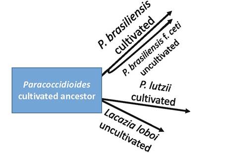
Fig. 9.2 The putative evolutionary paths of the culturable P. brasiliensis and P. lutzii recovered from humans with systemic paracoccidioidomycosis and the unculturable P. brasiliensis var. ceti and Lacazia loboi, restricted to subcutaneous tissues in dolphins and humans, respectively.
The diagram shows the phylogenetic placement of these pathogens according to current phylogenetic analyses with one or more genes (Vilela et al. 2009, 2016) (Fig. 9.1)One of the striking findings of the latter study (Vilela et al. 2016) was that after the addition of P. brasiliensis var. ceti, the DNA sequences of L. loboi showed lower bootstrap support to be in its own genus. This was best illustrated using the partial DNA sequences of the exons CHS4, gp43-like, and kex (Fig. 9.1) (Vilela et al. 2016). In previous analysis (Vilela et al. 2009), these exons placed the DNA sequences of human L. loboi with strong support as the sister taxon of two Paracoccidioides species (98-100% bootstrap support). However, with the addition of the dolphin DNA sequences, the bootstrap values plummeted to 83 (CHS4) and 48 (kex) (Fig. 9.1), and the gp43-like exon formed a strong supported sister taxon with P. lutzii, but did not affect bootstrap support using ITS DNA sequences. These analyses suggested that L. loboi maybe just another species in the genus Paracoccidioides (Vilela et al. 2016). The placement of the unculturable P. brasiliensis var. ceti among well-known DNA sequences of culturable isolates from human is intriguing. However, the fact that L. loboi and P. brasiliensis var. ceti shared many features strongly supports their common origin (Fig. 9.2).
9.3