Toxicologic Diseases
Numerous drugs, chemicals, plants, and fungal mycotoxŁins have been reported to cause toxic hepatitis specifically in goats. Recognition of toxic hepatitis and identification of the specific cause can be a difficult challenge to the practiŁtioner, requiring careful history taking, thorough environŁmental examination, complete physical examination, and often laboratory support.
The following discussion is divided into two parts. The first is a general discussion of the salient features of toxic hepatitis in goats. This is folŁlowed by brief descriptions of the specific known causes of the condition.General Features of Toxic Hepatitis
Diffuse destruction of liver parenchyma from various chemicals and drugs occurs sporadically in goats. Predisposing factors include overdosage of drugs, inapproŁpriate uses of drugs, idiosyncratic reactions, unsuspected exposures, and predispositions to toxicity.
Patterns of poisoning by plants are variable. In some cases, goats are offered poisonous plants unknowingly in the form of garden cuttings. Occasionally, poisonous plants are incorporated into hay, but not usually at concentrations capable of producing widespread toxicity. In grazing situaŁtions, the potential toxicity of plants may be altered by weather conditions or the occurrence of fungal parasites on plants that enhance toxicity. Many poisonous plants are unpalatable to goats under normal circumstances and will not be eaten readily. However, when drought or overgrazŁing occurs, goats may be compelled to eat toxic plants, and in fact may be more likely to eat them than are sheep and cattle due to their inherently diverse feeding behavior.
The development of hepatotoxicity is related to the dose of toxin, the action of the toxin, the duration of exposure, and the ability of the goat to detoxify the toxic agent. Drugs and chemicals tend to produce acute toxicosis after a single exposure, while most plant poisonings involve long-term feeding or grazing of the offending plant.
With some plants chronic disease develops gradually, while with others the plant toxicity is cumulative, and there is an extended period of normality before acute disease occurs.Biotransformation of toxic compounds to inactive metabolites occurs primarily in the liver and involves microsomal enzyme systems, particularly mixed-function oxidases, and the hemoprotein cytochrome P-450. Marked differences in various detoxifying activities occur among the ruminant livestock species (Patterson and Roberts 1970; Dalvi et al. 1987). Hepatic microsomal protein content of goats is equivalent to that of cattle, but significantly less than in sheep. The content of cytochrome P-450 is equivaŁlent to cattle but significantly more than in sheep, and goats have a significantly higher level of activity for the drug-metabolizing enzyme benzphetamine N-demethylase than either cattle or sheep. These findings suggest a basis for understanding the known variation in susceptibility to some toxic agents in goats, sheep, and cattle, and serve as a caution against presuming equivalent toxicities in goats for various xenobiotics based on their effects in sheep or cattle (Al-Qarawi and Ali 2003; Szotakova et al. 2004; Dacasto et al. 2005). Age can be a factor in biotransformation capacŁity as well. Livers of newborn goats exhibited very low activities of drug-metabolizing enzymes relative to 6-week-old and adult goats (Eltom et al. 1993).
In general terms, hepatotoxins manifest their toxicity by one or more of the following mechanisms: centrilobular necrosis, midzonal necrosis, periportal necrosis, cholestaŁsis, biliary hyperplasia, or venous occlusion. Acute, often fatal hepatic insufficiency can result when the initial toxic effects are severe. In less severe or chronic poisonings, lesions of ongoing hepatic necrosis are mixed with eviŁdence of attempts to heal, mainly characterized by fibrosis, leading over the long term to cirrhosis. Many of the hepa- totoxic agents, particularly plants, exert toxic effects in other organs as well, particularly the kidney, lung, and aliŁmentary tract when toxins are ingested.
The clinical findings in toxic hepatitis of goats vary with the cause. In general, depression and anorexia are the most consistent findings. Signs of hepatoencephalopathy, including weakness, ataxia, head pressing, torticollis, coma, and convulsions, are common. Evidence of liver pain, characterized by an arched back and resistance to liver palpation, are common in acute toxicoses. Jaundice is a less common finding than might be expected. Signs of photodermatitis may be observed in some plant poisonŁings. Because many hepatotoxic agents affect other organs as well, many clinical signs may be noted that are not hepatic in origin. Common findings in this category include dyspnea, diarrhea, and signs of renal failure.
In acute toxicity, serum levels of liver-associated enzymes are elevated. Liver-specific enzymes should be measured, because some toxic agents also damage muscle, causing increases in AST. Elevation of total bilirubin is not a conŁsistent finding in acute caprine hepatotoxicity. When elevations are observed, assessment of erythrocyte paramŁeters is indicated to determine if concurrent hemolysis is occurring.
Evaluation of liver function by BSP clearance and characterization of lesions by biopsy are helpful in conŁfirming the presence of toxic hepatitis in poisonings over the long term, because levels of liver-specific enzymes in the serum may not be pronounced in chronic disease. Anemia, hypoproteinemia, and hypoalbuminemia are common in chronic liver disease, but are by no means specific for hepatic dysfunction. When neurologic signs are observed, elevated blood ammonia levels are helpful in distinguishing hepatoencephalopathy from primary neurologic disease.
At necropsy, in acute toxic hepatitis the liver is swollen with rounded edges. On cut surface the lobular pattern of the parenchyma is accentuated by the presence of centriŁlobular necrosis and vascular congestion. When jaundice is present, organs and body fat appear yellow (Figure 11.5).
Edema, and less often ascites, may be seen when hypoproŁteinemia has had an opportunity to develop or when there is concurrent congestive heart failure. In many instances, the toxicity of hepatotoxic agents is not limited to the liver and multiple organ involvement is common. Gastroenteritis is common with ingested toxins and kidney lesions are freŁquent, presumably resulting from the toxin-filtering and concentrating action of the kidney. The rumen content should be examined for evidence of hepatotoxic plants.Definitive diagnosis of toxicity can be difficult. Success depends on careful history taking to identify all possible exposures to toxins and environmental examination to determine the presence of chemicals or plants unrecogŁnized by the animal caretaker. Although toxic principles of many poisonous plants are identifiable by laboratory
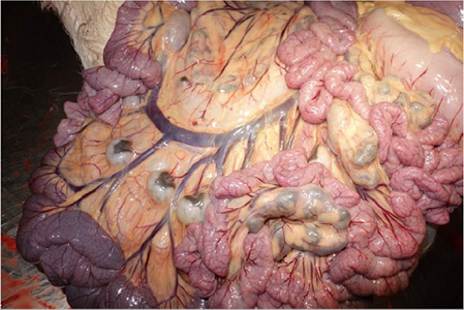
Figure 11.5 Icterus (jaundice) noted in the omental fat of a goat with hepatitis of unknown cause. Source: Reproduced by permission of Dr. Jaroslaw Kaba, Faculty of Veterinary Medicine, Warsaw University of Life Sciences, Warsaw, Poland.
methods, these methods are often expensive and not widely available for diagnostic use. Identifying the plant itself in the rumen content or feed or on grazing lands may be sufŁficient to make a presumptive diagnosis. The ability to identify fragments of toxic plants in rumen content is a valuable skill.
Specific treatments for toxic hepatitis are limited. Animals should be removed from risk of additional expoŁsure when possible. In known, acute oral toxicities, adminŁistering activated charcoal at a dose of 500 g for an adult goat may inhibit additional uptake of toxin from the gut. In individual valuable animals, rumenotomy might be attempted early in the course of disease to remove ingesta that may contain toxins. Supportive care includes fluids supplemented with glucose to counter dehydration and likely hypoglycemia, feeding low-protein diets to suppress additional hyperammonemia, and controlling seizures.
When photosensitization is present, animals should be kept out of direct sun, given corticosteroids to ameliorate the photodermatitis, and placed on antibiotics to prevent secondary pyodermas. In many cases of toxic hepatitis, aniŁmals that recover from acute disease may never return to full health or production potential due to permanent liver damage. The prognosis, therefore, should always be guarded.Controlling toxic hepatitis depends on identifying the toxic agent and preventing additional exposure. This includes accurate dosing of medications such as ivermecŁtin, substituting newer drugs for carbon tetrachloride in the treatment of liver flukes, restricting grazing of goats to avoid toxic plants, avoiding overgrazing, providing suppleŁmental feed during periods of drought, and recognizing periods of enhanced toxicity in certain plants. Obviously, accomplishing many of these goals is problematic. Additional recognition of environmental agents toxic to goats, particularly plants, and increased understanding of the patterns of occurrence of poisonings will lead to more specific recommendations for control.
Chemical and Drug-Related Causes of Toxic Hepatitis
Carbon Tetrachloride
Carbon tetrachloride administered as a flukicide can be hepatotoxic, even at the recommended dose of 1 mL/10 kg bw (Jones and Shah 1982). Overdosing can lead to acute death from respiratory arrest or a more prolonged three- to seven-day course involving hepatic and renal insufficiency. The sensitivity of goats to carbon tetrachloride toxicity was increased when animals were first treated with the chlorinŁated hydrocarbon insecticide dieldrin (Abdelsalam et al. 1982).
Hexachloroethane
Idiosyncratic reactions to routinely used drugs may occur, as reported in the case of Jamunapari goats in India given standard doses of hexachloroethane for treatment of liver flukes (Vihan 1987). Early signs of toxicity included rumen atony, bloat, diarrhea, and dyspnea, followed by depresŁsion, staggering gait, muscle spasms, and convulsions.
Necropsy lesions included hepatic necrosis, renal tubular degeneration, pulmonary edema, desquamation of foresŁtomach mucosa, and generalized hemorrhage. Such reacŁtions have also been reported in cattle and sheep.Halothane
Though no longer in common use in the United States, halŁothane gas anesthesia was used commonly in goats without incident. Nevertheless, there are two separate reports of presumed halothane toxicity producing massive hepatic necrosis with clinical signs of hepatoencephalopathy in the days immediately after anesthesia (Fetcher 1985; O'Brien et al. 1986). Although the exact mechanism remains unclear, it was postulated that a combination of prolonged anesthesia, hypoxia, and hypotension led to reductive rather than oxidative metabolism of halothane in the liver, resultŁing in production of hepatotoxic rather than inert metaboŁlites. In a subsequent study, careful monitoring of clinical enzymology in goats under halothane anesthesia for no more than two hours and histologic examination of liver samples one and two months later did not indicate any eviŁdence of liver damage or dysfunction (McEwen et al. 2000).
Iron
Iron toxicosis, caused by extralabel administration of an equine hematinic product to cattle, was reproduced experiŁmentally in goats given the same product (Ruhr et al. 1983). The product was formulated for intravenous use, but was given intramuscularly at approximately five times the recomŁmended horse dosage. Signs included respiratory distress, weakness, jaundice, recumbency, and convulsions. There was severe hepatic necrosis at necropsy.
Ivermectin
Ivermectin, given subcutaneously at 25 times the recomŁmended dose (5 mg/kg versus 0.2 mg/kg), produced acute collapse, hyperexcitability, and death, or a three- to four- day course of anorexia, weakness, recumbency, apparent blindness, coma, and death in Nubian goats. The principal postmortem lesion was a multifocal, non-suppurative, necrotizing hepatitis (Ali and Abu Samra 1987). In another study, East African goats were given eight times the recomŁmended dose and showed no adverse effects, except for some immediate, transient irritation at the subcutaneous injection site (Njanja et al. 1985).
Copper
Chronic copper toxicosis has been documented in young goats fed calf milk replacers containing copper (Belford and Raven 1986; Humphries et al. 1987). Chronic copper toxicity can cause hemolytic anemia as well as liver damŁage, though there has been a documented occurrence of chronic copper toxicosis in a dairy goat herd that showed no icterus or other signs of anemia (Cornish et al. 2007). Copper toxicity is discussed in more detail in Chapter 7.
Plants Causing Toxic Hepatitis
Caltrop
Geeldikkop, a hepatogenous photosensitization disease associated with grazing of T. terrestris (caltrop), a nutritious annual herb, cause severe economic loss in goat and sheep operations in South Africa (Kellerman et al. 1980). The conŁdition has also been reported from Australia (Glastonbury and Boal 1985; Jacob and Peet 1987), Iran (Amjadi et al. 1977), Argentina (Tapia et al. 1994), and California (McDonough et al. 1994). The plant, also known as puncŁture vine, yellow vine, and goathead, is found in warm, temŁperate, and tropical regions throughout the world. The disease occurrence is seasonal; the plant becomes toxic when wilted in hot, dry weather after summer rains. The hepatotoxicity and photosensitization are believed to be caused by steroidal saponins contained in the plant (Kellerman et al. 1991). Their toxic effect is enhanced when the plant is wilted. It has also been proposed that the presŁence of sporidesmin, a mycotoxin of Pithomyces chartarum, a fungus that grows on caltrops under the weather condiŁtions described, may enhance the toxic effects of the steroiŁdal saponins of T. terrestris. The clinical and pathologic effects of experimental poisoning of goats with T. terrestris have been described (Aslani et al. 2004).
Sacahuiste
Sacahuiste (N. texana) poisoning occurs in Texas and other portions of the southwestern United States only during a brief, three-week period in spring when this common range plant is bearing buds, blooms, or ripe fruit, because the leaves are not toxic. The period of blooming often coincides with a period of feed scarcity on rangeland, so consumption by goats, sheep, and cattle is more likely. Clinical signs include anorexia, depression, jaundice, dark yellow urine, purplish discoloration of the hoof, and photosensitization. The latter is more likely when green feed is available, because sacahu- iste blooms contain little chlorophyll (Mathews 1940a).
Lechuguilla
Agave Iecheguilla, or lechuguilla, is another important cause of caprine hepatogenous photosensitization in Texas, New Mexico, and northern Mexico during drought condiŁtions, especially in the spring. The disease is known locally as ōgoat feverö or ōswell-head.ö The plant is believed to contain two photosensitizing toxins, one of which is a sapŁonin. Signs include depression, loss of appetite, jaundice, yellow ocular and nasal discharge, photosensitization with swelling of the face and ears, and coma. Morbidity and mortality rates of 5-30% can occur (Mathews 1937).
Spurge
Poisoning by the spurge, Phyllanthus abnormis, occurs only in Texas in areas of sandy soil during drought periods when livestock eat young green plants. The toxin is degraded by drying. Goats are less susceptible than cattle, but can develop signs of listlessness, anorexia, unthriftiŁness, and diarrhea. There is severe fatty degeneration of the liver at necropsy (Mathews 1945).
Lantana
Lantana camara, which contains hepatotoxic lantadenes, is an important cause of poisoning in cattle and sheep in Australia, India, Mexico, and the United States. The shrub occurs commonly in many tropical and subtropical regions of the world and it may be consumed in large amounts durŁing periods of drought. The toxic syndrome is characterŁized by photosensitization, jaundice, rumen atony, and constipation. At necropsy, the carcass is icteric, the liver swollen and dark brown to black in color, and the gallbladŁder is distended. Enlarged kidneys and congested lungs may also be noted. Lantana poisoning with characteristic photosensitization has been reported to occur in goats in coastal eastern Australia (Seawright 1984). A naturally occurring case in a Boer kid was reported from South Africa in which there was icterus, dehydration, and constiŁpation, but no photosensitization (Ide and Tutt 1998). Two goats in India fed L. camara by their owner showed signs of anorexia and orange-colored urine within a day of ingesŁtion, and by the fourth day were dull and recumbent with jaundice and indications of photosensitization. Both recovŁered with supportive therapy (Chirayath et al. 2017). Attempts to reproduce the disease experimentally in goats caused hepatotoxicity with jaundice and some deaths, but no photosensitization in one study (Lin et al. 1985) and no clinical signs at all except loss of appetite in another (Lal and Kalra 1960). An experimental challenge conducted in India produced both jaundice and photosensitization (Ali et al. 1995). Additional documentation and description of field outbreaks in goats would be useful.
Pyrrolizidine Alkaloid Toxicity
In contrast with cattle and horses, but similarly to sheep, goats are relatively resistant to pyrrolizidine alkaloid toxicosis commonly associated with ingestion of Senecio spp., Crotalaria spp., and Heliotropium spp. of plants. This resistance is not absolute, but is dose dependent. Experimentally, signs and lesions typical of pyrrolizidine alkaloid toxicity were produced in goats by prolonged feedŁing of tansy ragwort (Senecio jacobaea) at total doses of 125-400% of bw, compared with lethal doses in horses and cattle of 5-20% of bw (Goeger et al. 1982b). Experimental challenge of goats with Crotalaria saltiana (Barri et al. 1984) and Heliotropium ovalifolium (Abu Damir et al. 1982) have also been reported. While toxicity was proŁduced in both cases, hepatic lesions consistent with pyr- rolizidine alkaloid toxicity, namely megalocytosis, biliary hyperplasia, portal fibrosis, and veno-occlusive disease, were absent or scarce. The toxic effects in goats challenged with these plants were presumed to be caused by toxic conŁstituents other than pyrrolizidine alkaloids.
It is unlikely that under normal grazing conditions goats would eat sufficient amounts of Senecio during the period of availability of the plant to produce severe, fatal, clinical disease. However, some hepatotoxicity can occur with chronic consumption and may impair growth and producŁtion. The use of goats and sheep to clear Senecio longilobus and Senecio spartoides from Texas rangeland to make it safe for cattle has been recommended (Dollahite 1972).
Even though goats may not become ill from long-term consumption of plants containing pyrrolizidine alkaloids, these hepatotoxins can be passed into the milk. Rats fed milk from goats receiving a ration containing 25% tansy ragwort developed liver lesions characteristic of pyr- rolizidine alkaloid toxicosis, though calves did not (Goeger et al. 1982a). Human poisoning by such a mechanism of relay toxicity from goat milk has not been reported.
Miscellaneous Plant Poisonings
A large body of research on plant poisonings in goats comes from the Sudan. The plants have been evaluated because they are commonly suspected to be involved in natural outbreaks of livestock poisoning, often under drought conditions. Plants found in the Sudan that were demonstrated experimentally to be hepatotoxic to goats include Heliotropium ovalifolium (Abu Damir et al. 1982), Crotalaria saltiana (Barri et al. 1984), Acanthospermum hispidum (Ali and Adam 1978), Jatropha aceroides, Jatropha glauca, Solanum dubium, Lagenaria siceraria, Citrullus colocynthis (Barri et al. 1983), Aristolochia bracte- ata, Cadaba rotundifolia (El Dirdiri et al. 1987), Ipomoea carnea (Abu Damir et al. 1987), Indigofera hochstetteri (Suliman et al. 1983), Capparis tomentosa (Ahmed and Adam 1980), and Tephrosia apollinea (Suliman et al. 1982).
In Brazil, extracts of the fruit of Sessea brasiliensis caused anorexia, incoordination, hyperexcitability, opisthotonos, and death in goats within 28-92 hours. Centrilobular necrosis and fatty degeneration of the liver were observed at necropsy. Vernonia mollissima, another Brazilian plant toxic to sheep and cattle, produced depression, staggering gait, recumbency with paddling, and death within 55 hours when fed to goats. Hepatic necrosis and renal damage were the predominant necropsy findings (Dobereiner et al. 1987).
In New Zealand, Vestiafoetida (Solanaceae) was reported to have caused severe periacinar necrosis and fatty change in the liver. Two young goats were observed consuming the plant. Following consumption the goats had clinical signs of mydriasis, loss of menace response, ataxia, muscle fas- ciculations, recumbency, and malodorous breath indicaŁtive of the crushed leaves of the plant. One responded to treatment with diazepam and vitamins and the other died. Necropsy revealed the liver lesions as well as fatty nephroŁsis (McKeough et al. 2005).
Hepatotoxicity has been observed in goats and cattle in Western Australia eating the leaves of Myoporum spp., including the Boobialla tree and Ellangowan poison bush. The toxic principle is a furanoid sesquiterpene essential oil that produces midzonal and centrilobular necrosis of the liver (Allen et al. 1978). Clinical signs include photosensitiŁzation, hemorrhage, dyspnea, and death.
Other sporadic causes of hepatogenous photosensitizaŁtion reported in goats include kleingrass (Panicum spp.) in the United States (Smith 1981) and signal grass (Brachiaria decumbens) in Nigeria (Opasina 1985) and Malaysia (Mazni et al. 1985).
The indolizidine alkaloids present in Oxytropis species can cause liver damage with vacuolation of liver observed histologically (Li et al. 2005). The clinical effects of Oxytropis spp. consumption are discussed in more detail in Chapter 5 relative to locoism.
Fungal and Mycotoxic Causes of Toxic Hepatitis
In South Africa, hepatotoxicity with photosensitization has been caused by a mycotoxin of the fungus Drechslera cam- panulata found growing on green oats (A. sativa) grazed by Boer goats. Clinical signs developed within eight days of the onset of grazing and included photodermatitis with edema of the head, diarrhea, and a 3.5% mortality rate (Schneider et al. 1985).
Aflatoxicosis has been reported in Sri Lanka in young goats receiving a concentrate supplement containing cocoŁnut meal contaminated with Aspergillus flavus. Clinical cases were observed over a seven-month period and the mortality rate was high. Jaundice was the most prominent sign, accompanied by lethargy, anorexia, a serous nasal discharge, and hypothermia terminally after a course of five to seven days. The livers were firm, congested, and fibrosed, and the gallbladder distended with tarry bile. There was biliary hyperplasia and periportal fibrosis microŁscopically. Similar findings have been reproduced experiŁmentally in goats (Samarajeewa et al. 1975; Miller et al. 1984). Decreased milk production has been demonŁstrated in aflatoxicosis and is correlated with the dosage of aflatoxin fed to goats (Hassan et al. 1985).
ōHard yellow liverö or hepatic fatty cirrhosis is a spoŁradic disease of grazing goats, sheep, cattle, and deer in west and south Texas causing chronic weight loss, neuroŁlogic signs, and terminal hepatic coma. At necropsy there is hepatic lipidosis and cirrhosis (Bailey 1985). The definiŁtive cause is unknown. It was hypothesized that a mycoŁtoxin, roridin A, extracted from Phomopsis fungus growing on grasses in endemic regions, was responsible for the disŁease (Samples et al. 1984). However, administration of purified roridin A to sheep for 60 days failed to produce liver lesions typical of the disease (Thormahlen et al. 1994), and the etiology remains unclear (Stegelmeier et al. 2019). The epizootiologic and clinical features of the condition have been reviewed (Helman et al. 1993).
The hepatic form of lupinosis, a mycotoxicosis associŁated with Diaporthe toxica (formerly Phomopsis leptostro- miformis) fungus on lupine plants, is an important disease of cattle and sheep in Europe, South Africa, and Australia. It has been produced experimentally in goats, but natural occurrence is poorly documented (Marasas 1974).