Traumatic Conditions
Predation
Predation can be a serious constraint on goat production. It is generally associated with death loss, particularly of neonates. However, non-fatal maiming of older goats occurs in certain situations, particularly those involving domestic dogs.
Predation is discussed in the present chapter because musculoskeletal trauma is a common outcome in these instances.Epidemiology
The subject of predator/prey relationships involving livestock is very complex and is fraught with economic, ecologic, ethical, and sociologic issues. For a more complete discussion of predation in general, the reader is referred to other sources (Gaafar et al. 1985; Rollins 2001; Shelton 2004).
Predation of goats is reported as a problem in Australia, the United States, and widely throughout the tropics. In Australia, predator importance varies with the type of goats involved and their geographic location, but is generally recognized as a major cause of kid mortality (P.J. Holst, Agricultural Research Station, Cowra, NSW, Australia, personal communication, 1986). Feral goat kids are lost to foxes, dingoes, feral pigs, and hunters. Milk goats, more likely kept in semirural or suburban areas, are preyed upon primarily by domestic dogs. Angora kids are lost to foxes, eagles, and urban and wild dogs. In Southern Australia, fox predation occurs in Cashmere goat flocks. Attacks are nocturnal and kids are the primary target. Nevertheless, kid losses caused by predation by foxes may be lower than those caused by mismothering and pregnancy wastage (Long et al. 1988).
In the United States, predation of hair goats and dairy goats has been studied. In south Texas, attacks on Angora goats were caused primarily by coyotes and secondarily by bobcats. Dramatic reductions in kid crop were attributable to predation, though aggressive predation control had little effect on reducing losses.
Adult goats were not preyed upon until kids were decimated. It was concluded that in areas of high coyote density, intense local predator control would be insufficient to prevent heavy kid losses (Guthery and Beasom 1978). In 1999, in Arizona, New Mexico, and Texas, three major goat-producing states, 61 000 goats and kids were lost to predators, with losses valued at $3.4 million (Howery and DeLiberto 2004). In the southern United States, migratory black vultures will also take goat kids.On 84 goat farms located in rural and semirural areas of Louisiana, domestic dogs were the most commonly recognized predators (Hagstad et al. 1987). Attacks occurred most commonly during periods of reduced light. Of goats attacked, 80% were killed outright or required slaughter, while 20% survived or recovered. Four management factors were identified as reducing the likelihood of attacks: penning of goats at night, the use of a night light, keeping goats near an occupied residence, and the presence of a dog belonging to the owner. Anecdotal reports from around the United States support the finding that domestic dogs allowed to run free at night are largely responsible for attacks on dairy goats. However, dairy goat owners in New England are increasingly complaining about attacks on kids by a growing coyote population.
Predation is cited as a cause of lost productivity throughout the tropics and housing goats at night is suggested as the most basic means of reducing losses (Devendra and Burns 1983). In a central Mali study, 17% of deaths in goats were ascribed to “lost” kids, presumably taken by predators. Uncontrolled domestic dogs and lax herding practices were considered responsible (Traore and Wilson 1988). In West Timor, Indonesia, goat herders identified predation by dogs and pigs as the third largest cause of kid death after navel infection and enteritis (Gatenby 1988).
Domestic dogs are more likely than wild predators to maim rather than kill goats, because the pursuit of goats begins more as a game than as an earnest attempt at food acquisition.
Domestic dogs often form packs that can wreak havoc in a flock or herd of goats with limited routes of escape. Far more goats are injured than could possibly be consumed as food. Goats and sheep make relatively easy targets for dogs because of their small size and non- aggressive behavior.Clinical Signs and Diagnosis
Evidence of predation is variable. Predation is often assumed when young kids are lost in areas where predators are endemic. Owners and herders may sometimes witness acts of predation or the presence of predators near flocks or herds. In the aftermath of an unobserved predator attack, evidence may suggest the type of predator involved (Guthery and Beasom 1978; Squires 1981; Long et al. 1988). Coyotes, for example, most often attack young kids and devour them completely. The presence of coyote spoor containing mohair in the vicinity of live - stock areas may be the only circumstantial evidence that coyotes are involved. Foxes may partially bury a carcass and, rather than devouring it completely, may slash open the abdomen and consume the entrails. When only small pieces of the carcass remain and there is wide scattering of blood, pigs are the likely predators. Two to four deep, focal penetrations of the skull, rib cage, or abdomen suggest killing by eagles. Primarily, predation must be differentiated from scavenging of animals dead of other causes. In predation extensive hemorrhage and free blood around the site of wounds occur, while in scavenging, such as that caused by crows, there is no hemorrhage and little free blood. Evidence of crow scavenging includes removal of eyes or tongue and evisceration through the anus or umbilicus.
In dog attacks, flesh wounds are common on the flanks and hindquarters, indicative of a chase, and carcasses may show no evidence of consumption. Animals surviving attack may show varying degrees of injury, from superficial skin and muscular trauma to severe trauma with extensive blood loss and shock. Injuries may include crushing fractures of bones and visceral damage caused by deep bites.
All bites can lead to secondary infection. While flesh wounds are relatively obvious, bites penetrating the abdomen and thorax should not be overlooked, due to the threat of secondary peritonitis or pyothorax.Treatment
The approach to management of goats after a predator attack depends a great deal on economics. When pet goats or valuable show or breeding stock are attacked and survive, aggressive medical therapy can be economically justified. Treatment of shock or blood loss with fluids, blood transfusion, and corticosteroid therapy is indicated. Broad-spectrum, parenteral antibiotic therapy should be initiated immediately and maintained for at least 10 days in cases of severe attack. Surgical interventions to manage visceral trauma may be immediately necessary, though orthopedic interventions can usually wait until patients are stabilized. If animals are brought for veterinary attention shortly after an attack, primary closure of skin and flesh wounds can be attempted. These should be left open or drains installed if it appears that much necrotic muscle will develop in wounds. Topical insecticide sprays should be used when fly strike is a potential problem. Affected animals should be kept in clean, warm, dry surroundings and fed a high-quality forage during convalescence. Tetanus prophylaxis is indicated. Non-steroidal antiinflammatory drugs or other analgesics should be administered as needed to alleviate the pain associated with traumatic injury, such as oral meloxicam at 1 mg/kg once a day, or intravenous or oral flunixin meglumine at 1.1-2.2 mg/kg once or twice a day. These drugs are reviewed in Chapter 17.
In commercial flocks, treatment of individual survivors may be economically constrained and the veterinarian should help the owner to minimize economic losses subsequent to dog attacks where large numbers of animals may be involved. Some suggestions made for the care of sheep flocks after predator attack are applicable to goats as well (Dille 1985).
It may be possible to shear hair goats and salvage the fleece. Hides may also be salable if not severely damaged. If fresh, carcasses may be sold as feed for animal consumption, for example to mink farms. Carcasses can also be used as bait to trap predators where trapping is allowed, or used as evidence to derive compensation from owners of dogs involved in attacks. There may be government compensation in some places for losses to predators.
In some circumstances, there may be an inclination to butcher for human consumption live animals that are seriously wounded and unlikely to recover. An abundance of caution should be exercised and this should not be done if there is any cause for suspecting rabies in the attacking animals, or if the affected animal has an elevated or subnormal temperature. If butchering is carried out, areas around bite wounds should be liberally trimmed. Animals unable to walk after attack and those with evidence of penetration of body cavities by bite wounds should only be considered for emergency slaughter if the bite wounds are fresh.
Control
At the flock or herd level, losses to predation may be controlled in a number of ways. Active destruction of predators through trapping, poisoning, or shooting may be subject to local regulation and may or may not be economically justified when the costs of predator control are weighed against losses. Each situation should be evaluated on its merits. When domestic dogs are involved, stock owners must be sure that local ordinances controlling unleashed dogs are adequate and rigidly enforced. Poison baits for controlling predators have been withdrawn from use in the United States since 1972 and other methods, such as trapping, snaring, M-44 sodium cyanide ejectors, gas cartridges for denning coyotes, and livestock protection collars, have been restricted or eliminated by ballot initiatives in some states (Andelt 2004).
As predator control options have become more restricted, the use of livestock-guarding animals has emerged as a cost-effective and humane approach to protecting small ruminants from predation and their use has been reviewed (Andelt 2004).
A number of dog breeds selectively developed in Europe and Asia to protect livestock from bears and wolves have become popular as guarding dogs for small ruminant flocks in the United States. These include the Great Pyrenees, Akbash, Anatolian Shepherd, and Komondor breeds, as well as the Maremma and Shar Planinetz breeds. Guard dogs in the United States are reported to be effective against coyotes, black bears, grizzly bears, and mountain lions, but may not be effective against wolves (Andelt 2004). Donkeys and llamas are also used as herd guardians, with the advantage that they eat what the goats eat and thus do not require separate feeding as do dogs. Some donkeys will attack and maim goats, so they should be closely observed when initially introduced. Llamas are particularly effective against dogs, coyotes, and foxes, because they are naturally aggressive toward canids. Intact males may try to breed ewes, so female llamas or geldings are preferred. Fencing is a costly and often inefficient means of predator control, though in Australia dingo-proof fencing has been helpful to sheep farmers.At a minimum, where predators are a problem, surveillance of the flock should be increased during kidding times and goats confined to a restricted kidding area when management conditions permit. Goats should be kept in a pen at night and the holding area should be illuminated. Control is improved if the holding area is near an occupied residence. Placing a baby monitor in the goat pen and in the owner's home may help alert owners to disturbances associated with predators. Where migratory black vultures inflict serious losses on kids, it may be necessary to adjust the breeding season to finish kidding before the vultures arrive in the spring.
Fractures
Fractures are common in goats, though epidemiologic data on prevalence and predisposing factors is limited.
Epidemiology
Environmental hazards are often implicated in the occurrence of fractures. All forms of gating, fencing, and penning material should be evaluated carefully before the material is used for the likelihood of goats catching limbs or necks in structural spaces. The curiosity and climbing instincts of goats ensure that they will find a way to become entangled whenever possible. In the author's (DMS) experience, chain-link fences especially predispose to limb fractures in intensively managed goats. The animals stick their legs through the fence and then, if frightened or crowded by other goats, are unable to extricate the limb. Fracture secondary to struggling is a likely outcome. In browsing goats, a leg may be caught in the fork of a tree, with similar consequences (Figure 4.14). Trauma from dog attacks is another common cause of limb bone fractures in goats in the United States. Preexisting bone diseases such as osteomyelitis, chronic fluorosis, fibrous osteodystrophy, osteoporosis, or rickets can predispose to fractures.
Two surveys from India give differing perspectives on the occurrence of caprine fractures. In Hisar, most fractures occurred in goats between 1 and 3 years of age. In order of decreasing frequency, fractures were observed in femur, tibia, metacarpus or metatarsus, phalanx, humerus, radius, and ulna (Singh et al. 1983). The prevalence of femoral fractures was attributed to free-ranging goats around villages being hit from behind by vehicles with bumpers at thigh level. In Ranchi, fractures occurred most often in goats younger than 6 months of age. The bones most often involved were, in descending order of frequency, metacarpus, metatarsus, femur, tibia, radius and ulna, humerus, and phalanx. Forelimb and hindlimb fractures occurred
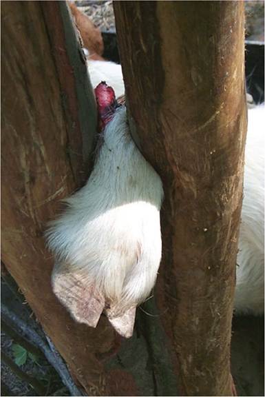
Figure 4.14 This goat caught its forelimb in the fork of a tree at pasture and in struggling to free itself, suffered an open fracture of the trapped limb. The goat was discovered alive and was euthanized. Source: Courtesy of Dr. M.C. Smith.
heal. The reader is referred to textbooks of large animal surgery for discussions of surgical repair of fractures. Suffice it to say here that numerous approaches to successful fracture management have been reported in goats, including compression plating of radial and ulnar fractures (Bacher and Potkay 1975) and full limb casting of tibial fractures, followed by application of a Robert Jones bandage (Mbiuki and Byagagaire 1984). Use of plaster casting alone was found to be an unsatisfactory method of managing radial and ulnar fractures (Buchoo and Sahay 1987).
When fracture repair and return to normal function are complicated by osteomyelitis or permanent nerve damage, limb amputation has been demonstrated to be a reasonable salvage operation in goats (Misk and Hifny 1979; Gamsjaeger and Chigerwe 2018). The pelvic limb can be disarticulated at the hip or stifle or amputated at the midfemoral level. Treated goats show excellent adaptation to three-legged locomotion if they are not allowed to become overweight.
Control
When fractures occur more than sporadically in a herd or flock, underlying problems of structural hazards or nutritional deficiencies must be considered as predisposing to an increased incidence of skeletal trauma. Careful attention must be paid to calcium, phosphorus, and vitamin D levels in the ration.
with approximately equal frequency. The occurrence of fractures was believed to be predisposed by soil calcium and phosphorus deficiencies and the hilly terrain in the region. Causes of fracture identified by history included slipping while grazing on hillsides (54%), playing or gamboling (24%), automobile accidents (18%), and trapping of limbs in wire enclosures (4%) (Dass et al. 1985).
Clinical Signs and Diagnosis
Fractures are usually identifiable by an acute onset of lameness, deviation of limbs, and/or palpable crepitus. Fractures secondary to nutritional deficiency rather than trauma are less likely to displace or be recognized on physical examination. Radiographic studies help to define the nature of the fracture and suggest therapeutic approaches.
Treatment
In young kids, simple splinting of distal limb fractures is frequently adequate and economically advantageous. Toothbrushes and split sections of PVC pipe are examples of splints that permit closed fractures of young kids to heal within three weeks. Adults may require five to six weeks to