Upper Respiratory Tract Diseases
As discussed earlier, clinical signs such as stertor, sneezing, nasal discharge, and cough suggest but are not limited to conditions with upper respiratory tract involvement.
Rhinitis
There are several possible etiologies for inflammation of the nasal passages.
Irritation from foreign material or parasites should be ruled out before more esoteric explanations are sought. Two mature goats in Brazil developed Protot⅛ecα-induced pyogranulomatous lesions of the skin at the nares and extending onto the nasal mucosa, causing inspiratory stertor, dyspnea, and weight loss (Macedo et al. 2008; Camboim et al. 2011). Granulomatous fungal rhinitis due to conidiobolomycosis (do Carmo et al. 2020) and aspergillosis (do Carmo et al. 2014) has also been seen. These reports underscore the importance of obtaining a biopsy sample from perplexing cases.Caprine Herpesvirus
Caprine herpesvirus infection (see Chapter 12) has caused severe generalized signs, including bloody diarrhea, fever, dyspnea, and purulent nasal discharge. Fibrinonecrotic ulceration of the nasal septum occurred in one kid experimentally infected (Berrios et al. 1975). In other experimental infections, the virus has caused a catarrhal rhinitis and mild tracheitis (Buddle et al. 1990a).
Nasal Bots
Nasal bots (Oestrus ovis) cause a chronic catarrhal to purulent discharge (Prein 1938) that may contain many eosinophils. This parasite is less common (larval burdens are smaller) in goats than in sheep (Dorchies et al. 1998), although one study from Iran documented a 13% prevalence in goats at slaughter (Shoorijeh et al. 2011). The adult fly deposits larvae around the nostrils of animals on pasture. First instars enter the nasal cavity and develop into second instars, which invade the sinuses of the head. Mature larvae return to the nostrils (if they have not grown too large to escape from the sinuses) after 2-10 months and are expelled by sneezing (Kimberling 1988).
Pupation occurs in the ground. Adult flies emerge after four weeks or longer, or possibly the next spring. Occasionally larvae are deposited about the eyes of humans, causing painful conjunctival ophthalmomyiasis (Cameron et al. 1991).Nose bots in goats can be suspected if nasal discharge is profuse and contains eosinophils. Frequent violent sneezing and avoidance behavior in late summer are typical. Caked-on dust leads to mouth breathing and interference with feeding, while inhaled bacteria and eosinophils may induce lung damage. Repeated infections induce a hypersensitivity and immunity in goats (Dorchies et al. 1998). Sometimes the bots are just an incidental finding in the sinus upon dehorning of adult goats.
The condition can usually be ignored if it does not interfere with feed consumption. Various questionable products have been sprayed into the nasal passages in late fall and winter to kill larvae, including Ruelene® aerosol (Dow Chemical Co., Midland, MI, USA) and concentrated Lysol® (Reckitt Benckiser, Parsippany, NJ, USA) diluted 1 : 5 with water. Ivermectin (even at 0.2 mg/kg level) and moxidectin are highly effective against all stages of nose bots. A prolonged milk withdrawal of nine days is suggested if ivermectin is administered orally to goats producing milk for human consumption, and 40 days if given subcutaneously (SC) (Baynes et al. 2000). Eprinomectin pour-on at 0.5 mg/ kg has been shown to be effective against O. ovis in sheep (Hoste et al. 2004) and is less likely to contaminate milk.
Nasal Leech
The nasal leech (Dinobdella ferox) infests ruminants grazing at altitudes of 900-1800 m in the foothills of the Himalayas. The leeches live in permanent pools and springs; young leeches attach themselves to the nostrils of animals that drink there during the dry season. Leeches migrate to the nasal passages to feed, but drop back into the water at the onset of the monsoon season (mid-June). Clinical signs include sneezing, blood-tinged mucoid nasal exudate, intermittent epistaxis, and anemia (Mahato et al.
1993). When leeches become large enough to protrude through the nostrils (15 days after infection) and restrict breathing, goats become restless and anorectic. Treatment is achieved by wetting the goat's nostrils with water to encourage leeches to protrude, then permitting the leeches to dip their bodies in a (reusable) solution of 10 μg∕mL ivermectin. Leeches are expelled within a few hours after this therapy. Systemic ivermectin is not effective (Mahato 1989).Nasal Schistosomosis
The blood fluke Schistosoma nasale has caused nasal obstruction (“snoring disease”) in goats in India. This parasite is discussed in Chapter 8.
Foreign Matter
Foreign body rhinitis is often caused by powdery feeds. Regurgitation is a more serious possible cause. Assuming the absence of a cleft palate, nutritional muscular dystrophy (white muscle disease) should be considered whenever milk runs out of a kid's nose. This is discussed under inhalation pneumonia. Vagal nerve irritation in the abdomen or thorax or poisoning by plants of the Ericaceae (rhododendron) family may cause regurgitation through the nose and mouth, but the distinctive smell and green color of rumen contents on the muzzle make the identification of the irritating substance simple.
Bacterial Rhinitis
Atrophic rhinitis associated with toxigenic strains of Pasteurella multocida has been recognized in goats in Norway (Baalsrud 1987). Signs observed in affected herds include purulent nasal discharge, nose bleeding, sneezing, and occasionally tender or distorted noses. Turbinate atrophy can be recognized when heads are sectioned at the level of the first premolar teeth. In tropical countries, purulent rhinitis with raised coalescing nodules on the nasal septum and turbinates has been observed in goats with melioidosis (Omar 1963). This infection is discussed under bacterial pneumonia below.
Neoplasia
An enzootic nasal tumor of young adult goats has been reported from France and Spain and recognized in individual animals elsewhere in the world (Fontaine et al.
1983; Pringle et al. 1989; De las Heras et al. 1991a, 2003b). A large outbreak (41 cases in goats 6 months to 6 years old) occurred in Italy after the importation of Alpine goats from France (Vitellozzi et al. 1993). The lesion is unilateral or bilateral and papillary and results in lysis of the turbinates. Histologically, the tumor is benign. Retroviral-like particles have been demonstrated in the tumors of several goats that had negative agar gel immunodiffusion tests for maedi- visna (and therefore CAE) virus (De las Heras et al. 1988, 1991a). Nasal secretions and the associated type D retroviral particles (now classified as a beta retrovirus, ENTV-2) have transmitted the tumor experimentally (De las Heras et al. 1991b, 1995). Affected animals show profuse seromucous nasal exudate, coughing, dyspnea, and stertor. One or both eyes may protrude and pressure on softened cranial bones causes expulsion of mucous nasal exudate. The outcome is eventually fatal, the result of emaciation and asphyxiation (De las Heras et al. 1991a). Presumably, select cases might be amenable to surgical extirpation, as has been performed for nasal tumors of sheep (Rings and Rojko 1985; Trent et al. 1988), but recurrence would be expected due to the viral etiology. An antemortem test using reverse transcription (RT)-PCR on RNA extracted from nasal swabs has been developed for the closely related ENTV-1 virus of sheep (Walsh et al. 2014). Serologic tests did not correlate well with disease status.Lymphosarcoma can also invade the nasal sinuses of goats (Craig et al. 1986). A fungal granuloma caused by Cryptococcus neoformans has been reported to obstruct the nasal cavity of a goat in Australia (Chapman et al. 1990) and could be easily mistaken for a tumor.
Traumatic Pharyngitis
Balling gun injuries can cause severe cellulitis in the region of the pharynx. Small plastic balling guns are particularly dangerous, because the goats soon make them very rough by chewing on the plastic when bolus administration is attempted.
Common clinical signs include cough, nasal discharge, and secondary inhalation pneumonia. The breath may have a foul odor, and the region of the pharynx is swollen and sensitive to palpation. Examination is simplified by tranquilization. Broad-spectrum antibiotics are given for treatment. Prevention typically involves using an alternative to anthelmintic or sulfonamide boluses.Abscessation of Retropharyngeal
Lymph Nodes
An enlarged retropharyngeal lymph node (Figure 9.3) may cause stertor, dyspnea, and coughing because of pressure on the pharynx or trachea. The mass can be detected by careful palpation and by radiography or even computed tomography (Grosso et al. 2018). The abscess is often caused by Corynebacterium pseudotuberculosis. A brave and careful surgeon is required to marsupialize the abscess without severing carotid artery, vagus nerve, or other important structures. An alternative approach described in a ram involves passing a flexible endoscope through one nasal passage and
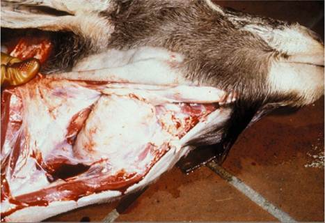
Figure 9.3 Abscessation of the retropharyngeal lymph node caused marked dyspnea in this goat. Source: Courtesy of Dr. J.M. King.
using a laser to lance the abscess into the pharynx (Copeland et al. 2020). Placement of a cuffed tracheostomy tube under local anesthesia has been recommended before induction of general anesthesia (Benson 1986). Caseous lymphadenitis is discussed in detail in Chapter 3.
Laryngitis or Tracheitis
These conditions are suspected when stridors are audible and can be localized to the corresponding portions of the upper respiratory tract. As discussed earlier, the trachea of a normal goat feels quite small.
Common Causes
Collars that press on the trachea may induce coughing or even damage the trachea in a tethered goat or one that is led by a collar. The cough may be incidental, but the danger of leg injury to the goat from a tether is very real.
Tracheitis because of irritants such as smoke, dust, whitewash, and ammonia in urine-soaked bedding has already been alluded to in the discussion of coughing. The diagnostician should get down to goat level and inhale deeply. If hay is dusty, the owner should try shaking it out while the goats are out of stall. If a goat is eating well and maintaining normal production and body condition, occasional coughing when fed or aroused is of no real concern.The possible role of Mycoplasma ovipneumoniae (see below) in chronic cough of otherwise healthy kids on certain farms remains to be studied. This cough is usually outgrown by about 8 months of age. In lambs, this syndrome has been associated with autoantibodies against respiratory cilia, possibly induced by M. ovipneumoniae infection (Niang et al. 1998b).
A single case of laryngeal hemiplegia has been reported in an adult Alpine goat (Tschuor et al. 2007). No etiology was determined, even with full necropsy and evaluation of the (normal) laryngeal nerve and musculature. In northeastern Brazil, stridor associated with degeneration of the recurrent laryngeal nerve and atrophy of the vocal folds and laryngeal muscles has been observed in goats with severe copper deficiency (Sousa et al. 2017).
Necrotic Laryngitis
Fusobacterium necrophorum is a sporadic cause of laryngitis and of necrotic stomatitis in goats. The condition is discussed in Chapter 10.
Cilia-Associated Respiratory Bacillus
A Gram-negative filamentous bacterium called cilia- associated respiratory bacillus, or CAR, has been associated with respiratory tract disease in laboratory rodents, cattle, deer, and pigs, among other species. This organism has also been found by histologic examination (Warthin- Starry stain and immunohistochemistry) and electron microscopy in the trachea of kids and adult goats with chronic tracheitis (Fernandez et al. 1996; Oros et al. 1997a) and in the lungs of slaughtered goats with enzootic pneumonia (Oros et al. 1997b). The bacteria are interspersed with cilia and oriented perpendicular to the surface of the tracheal and bronchial epithelium. The significance of CAR in goats remains to be determined and there has been little research on the topic in recent years.
Infectious Bovine Rhinotracheitis
Infectious bovine rhinotracheitis (IBR) virus (bovine herpesvirus type I) is rarely isolated from goats, but experimentally causes pyrexia and mild clinical signs. Cough and nasal discharge would be expected if any signs at all occurred, although there is one report of severe respiratory disease (Mohanty 1972). The use of IBR vaccination is not recommended for goats. In fact, several caprine isolates have been indistinguishable from bovine vaccine strains (Whetstone and Evermann 1988). Some Mediterranean countries report a high seroprevalence of bovine herpesvirus type 1 in goats in the absence of vaccination programs (Mahmoud and Ahmed 2009). Goats may be latent carriers of the virulent bovine virus, potentially interfering with eradication attempts in cattle herds (Six et al. 2001; Yesilbag et al. 2003). Goats are also susceptible experimentally to acute and latent infection by bovine herpesvirus type 5, showing mild respiratory signs and conjunctivitis (Diel et al. 2007).