Viral Causes of Pneumonia
Viral pneumonias in goats can be chronic or acute. Secondary bacterial infections are often superimposed.
Respiratory Syncytial Virus
A single caprine isolate of respiratory syncytial virus (RSV, a single-stranded RNA pneumovirus of the family Paramyxoviridae, subfamily Pneumovirinae) has been characterized in the United States (Lehmkuhl et al.
1980). This isolate is very closely related to strains of bovine respiratory syncytial virus (BRSV) (Trudel et al. 1989). An RSV similar to BRSV has been isolated from goat kids with respiratory disease in Spain (Redondo et al. 1994). Clinical signs reported in this outbreak included fever, loss of appetite, conjunctivitis, nasal discharge, cough, and tachypnea. Animals with a simultaneous fibrinous Mannheimia haemo- lytica bronchopneumonia had higher fevers and more labored breathing. Crackles were heard over the lungs of all affected animals. Histologic lesions included diffuse interstitial pneumonia and syncytia formation.Several surveys have documented antibodies against this virus or the closely related bovine RSV in healthy goats. This includes 36% of 112 adult goats from 40 herds studied in Quebec (Lamontagne et al. 1985), 31% of 318 goats in 22 herds in another Quebec study (Elazhary et al. 1984), 50% of 332 goats in Louisiana (Fulton et al. 1982), and 11 of 40 goats from farms in the Netherlands that also harbored cattle (Van der Poel et al. 1995). Goats in England were found to have antibodies against BRSV without ever having shown respiratory signs (Morgan et al. 1985), whereas goats seroconverted during a pneumonia outbreak in Zaire (Jetteur et al. 1989).
The importance of RSV as a respiratory pathogen in goats remains unclear. However, practitioners in the United States report that the virus is frequently demonstrated (by virus identification or seroconversion) in goats that develop a fever and respiratory signs after a show.
Treatment with ceftiofur may be administered for possible concomitant bacterial infections, which cannot be ruled out until laboratory test results become available. There are no publications available concerning benefits ofvaccination, although some veterinarians have used univalent BRSV vaccines to prevent respiratory disease in show goats. There are no studies documenting efficacy of these vaccines in goats.
Progressive Interstitial Retroviral Pneumonia
Retroviruses (lentiviruses) are believed to cause both sub- clinical and fatal pneumonia in goats.
Etiology
The caprine arthritis encephalitis virus (CAEV, viral leu- koencephalomyelitis) has been reported to cause subclini- cal interstitial pneumonia (Cork et al. 1974). Occasional kids with neurologic lesions also have diffuse involvement of one or more lung lobes.
A clinical syndrome indistinguishable from ovine progressive pneumonia and maedi-visna has been identified in CAEV-infected adult goats (Robinson 1981; Oliver et al. 1982). The sheep and goat viruses are considered to be distinct by most researchers, but serologic tests cross-react and both viruses are spread through milk, as discussed in Chapter 4.
To date it has not been possible to reproduce this condition experimentally. Therefore there is a suspicion that an additional agent, such as a helper virus or a bacterial infection that activates viral-infected macrophages, may be involved. There is currently no evidence that the production of interstitial pneumonia depends on the strain of CAEV with which the goat is infected (Ellis et al. 1988b). Inoculation of CAEV into Merino lambs has failed to produce clinical disease in the sheep (Dickson and Ellis 1989). Natural transmission of small ruminant lentiviruses between sheep and goats has been documented in mixed herds (Shah et al. 2004).
Clinical Signs
The first signs of dyspnea usually occur after a stress such as kidding or mastitis. The clinical course may be several weeks in advanced pregnancy or many months in the doe first affected after kidding.
Exercise intolerance, dyspnea, and wasting often accompanied by a cough are prominent. Some but not all affected goats have enlarged carpi. Secondary bacterial pneumonias may be superimposed or may have been the trigger for the interstitial pneumonia, and antibiotics often appear to give temporary relief. The pulmonary form of caseous lymphadenitis has a similar clinical appearance.Diagnosis
Radiographs show small patches of interstitial pneumonia early in the course. A more advanced case is depicted in Figure 9.4 and elsewhere (Koenig et al. 1990). As debilitation progresses, large areas of lung are consolidated and bullous emphysema may damage the remaining lung parenchyma. Either caudal lung lobes or cranioventral lung lobes may be involved (Ellis et al. 1988a). At necropsy, the affected areas are swollen, gray-pink, and firm (Figure 9.5); numerous 1-2 mm whitish gray foci are visible on cut section, as shown in Figure 9.6 (Robinson and Ellis 1984). Mediastinal lymph nodes are enlarged. Lung biopsies might confirm the diagnosis before death. Serologic testing is of limited diagnostic value, but should identify an infected herd.
Histologic study of caprine progressive pneumonia reveals interstitial (peribronchiolar) accumulation of mononuclear cells and proliferation of type II pneumocytes. Alveoli
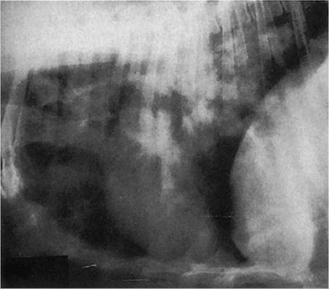
Figure 9.4 Pulmonary radiograph of a goat with interstitial pneumonia. Source: Courtesy of Dr. M.C. Smith.
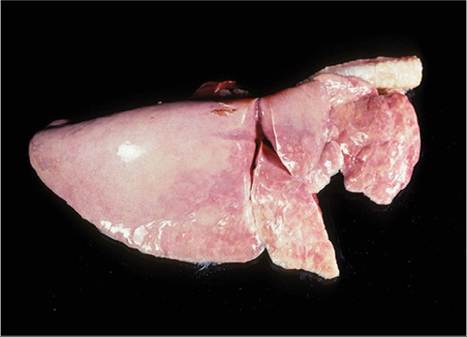
Figure 9.5 Goat lung with caprine arthritis encephalitis interstitial pneumonia distending the diaphragmatic lobe. Source: Courtesy of Dr. M.C. Smith.
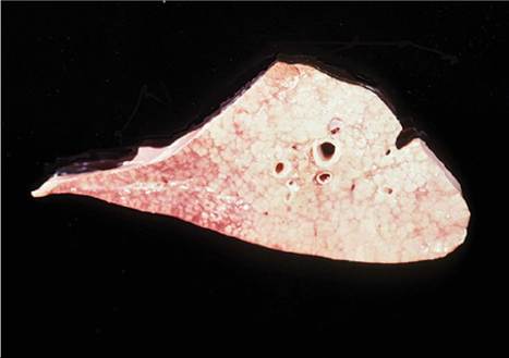
Figure 9.6 Cross - section of an affected lung lobe, showing infiltration of the pulmonary parenchyma. Source: Courtesy of Dr.
M.C. Smith.are filled with an eosinophilic material that resembles surfactant when examined with the electron microscope (Robinson and Ellis 1984, 1986).
Discussions of the other CAEV-associated syndromes (i.e., arthritis, neurologic disease, mastitis) can be found elsewhere in this text. Control procedures are discussed in detail in Chapter 4.
Ovine Pulmonary Adenocarcinoma, Sheep Pulmonary Adenomatosis, Jaagsiekte
This contagious lung tumor of sheep has been reported infrequently in goats (Rajya and Singh 1964; Stefanou et al. 1975). The disease has been reported in sheep on all continents except Australia. The cause is an oncogenic beta retrovirus (De las Heras et al. 2003a). Transmission appears to be via respiratory secretions. Experimental infection is much more difficult to produce in goats than in sheep (Sharp et al. 1986; Tustin et al. 1988).
Clinically affected animals are mature and afebrile (in the absence of secondary bacterial infections) and show prolonged weight loss and dyspnea. Fluid accumulates in the lungs, which results in auscultable crackles or rales. A simple test used to differentiate the condition from chronic progressive pneumonia is to elevate the animal's hindquarters. Fluid is expected to run out the nose if the animal is severely affected with adenomatosis. The disease is considered to be invariably progressive and fatal. There is no known treatment and no serologic test. Control is usually limited to culling goats that are wasting, although a PCR test for proviral DNA in leukocytes has permitted identification of subclinically infected sheep (Gonzalez et al. 2001). Removal at birth and artificial rearing on colostrum substitutes and milk replacer (essentially a CAE eradication program) has also been successful in creating a disease-free sheep flock (Voigt et al. 2007).
At necropsy, many grayish nodules or extensive solid tumors are found in the lungs. The respiratory passages are filled with white froth.
Histologically, there are alveolar and intrabronchiolar growths composed of masses of cuboidal and columnar cells. Metastases to regional lymph nodes have not been reported in goats (Sharma et al. 1975), but there is one report of metastasis to heart and kidney (Al-Dubaib 2005).Peste des Petits Ruminants
This important infectious disease of sheep and goats is also called PPR, stomatitis pneumoenteritis complex, and Kata (Hamdy et al. 1976).
Etiology and Pathogenesis
The etiologic agent is a paramyxovirus of the Morbillivirus genus that is not pathogenic for cattle. The closely related rinderpest virus of cattle caused similar signs in goats before it was eradicated from the world (Roeder et al. 2013). The peste des petits ruminants (PPR) virus seriously limits goat production in West Africa, the Middle East, India, and Southeast Asia.
The virus is shed in secretions to infect other animals by direct contact. It can persist in the environment for as long as 36 hours, and thus holding pens can be a source of infection. The disease is discussed in detail in Chapter 10.
Clinical Signs
The disease is characterized by fever persisting for five to seven days and a profuse, even bloody diarrhea. Necrotic stomatitis, foaming at the mouth, and an ocular discharge are also noted. Pregnant animals may abort (Abu Elzein et al. 1990). Respiratory signs occur during the acute stages; these include malodorous mucopurulent nasal discharge, frequent sneezing, increased respiratory rate, extended head, and mouth breathing. Severe leukopenia persists for 10 days in animals that recover (Scott 1990). Some goats die two to three weeks later of a secondary bronchopneumonia (Whitney et al. 1967). The highest incidence of disease occurs during the rainy season, and goats most commonly affected in enzootic regions are 6-12 months of age. Kids younger than 3 months of age usually have colostral immunity. Adults will also be affected in naive populations.
Diagnosis
Fluorescent antibody testing was previously used for identifying the virus in nasal discharges and intestinal scrapings.
When available, an RT-PCR test is now used to identify the virus in oculonasal swabs, oral lesions, or blood (Qam et al. 2005). More recently developed tests for the virus have been reviewed by Banyard et al. (2010) and are discussed in Chapter 10. A pen-side test has been developed using a lateral flow device to detect viral antigen in ocular and nasal swabs (Baron et al. 2014). This test permits differentiation of PPR from simple bacterial pneumonia in live goats in situations where sophisticated laboratory facilities are not available.The necropsy findings relative to the gastrointestinal tract are described in Chapter 10. There may be consolidation of cranioventral lung lobes. Histologic lesions reported in the lung include a giant cell pneumonia, with eosinophilic intracytoplasmic inclusions in epithelial cells in perhaps half of the goats dying because of PPR. Less commonly there is necrosis of the tracheal epithelium. Diagnosis is complicated by secondary Pasteurella, Mannheimia, or Mycoplasma pneumonia.
Prophylaxis
Vaccination against this disease in endemic areas is both feasible (Gibbs et al. 1979) and economically sound (Opasina and Putt 1985). Vaccination is central to the World Health Organization for Animal Health's plan for global eradication of the virus (Cameron 2019). Prophylaxis is covered in Chapter 10.
Hyperimmune serum (5 mL IV) reverses the signs of PPR if given during the febrile stage of the disease, when the temperature is 40.5 °C (104.9 °F) or higher. However, reinfection or relapse occurs after 10 days, and even those goats that develop labial scabs and appear to recover are susceptible to later challenge (Ihemelandu et al. 1985). Thus, goats treated with hyperimmune serum should still be vaccinated. In the context of the global eradication effort that has begun, treatment of individual animals infected with PPR virus may not be permitted in the future.
Goat Pox
The capripox virus, discussed in detail in Chapter 2, causes systemic signs that include pyrexia, anorexia, and an arched back. Case fatality rates are increased when the respiratory tract is involved, and young goats are more severely affected than adults (Rao and Bandyopadhya 2000). At necropsy, multiple foci of consolidation (0.5-2 cm diameter) are commonly found beneath the pleura. These lesions may be hemorrhagic (Kitching 2004). Similar lesions may be seen in other organs such as liver, kidney, and abomasum. Secondary bacterial pneumonia is commonly the cause of death (Davies 1981).