AVIAN RESPIRATION
Aside from subtle differences, many basic features of respiration in mammals apply to birds (e.g., respiratory pressures, oxygen transport, carbon dioxide transport, regulation of respiration).
The description of avian respiration that follows assumes familiarity with basic features and addresses major differences. Major differences will be noted in respiratory morphology and the mechanics of respiration and air circulation.General Scheme of Avian Respiratory Morphology
1. Do avian lungs expand and contract during respiratory cycles?
2. Are air sacs part of the lungs?
3. Do air sacs expand and contract during respiratory cycles?
4. What is the more common name for tertiary bronchi?
5. What composes the parabronchial mantle?
6. What part of the avian lung accounts for gas exchange?
7. How many air sacs are there? Where are they located?
8. Do the air sacs account for significant gas exchange?
9. Where would blood perfusion be more abundant, in the air sacs or the lungs?
0. Could smoke enter a broken wing bone (humerus) and exit the trachea?
There are important physiological differences between wild birds and domesticated birds. In this regard, the study of avian physiology that follows will be related to domesticated birds, and more particularly to the chicken. The respiratory apparatus,of birds is decidedly different than that of mammals. The organ of phonation, the syrinx, is located at the bifurcation of the trachea, near the lungs, rather than the larynx. Also, the tracheal rings are complete, rather than incomplete as in mammals. Beyond the trachea, more striking differences are apparent. The lungs continue to be the gas exchange structures, but they do not expand and contract during respiratory cycles. They are relatively small and are fixed in position by their attachment to the ribs. Their ventilation depends on bellows-like extensions from the lungs known as air sacs, which do expand and contract during respiratory cycles, as will be described later.
The lungs and air sacs are served by airway divisions from the trachea known as primary, secondary, and tertiary bronchi. The tertiary bronchi are more commonly known as parabronchi. There are two sets of parabronchi and they are known as paleopulmonic parabronchi and neopulmonic parabronchi. The latter set is caudle to the former and exists just cranial to the caudle air sacs. The relationship of the bronchi to each other and to the air sacs is shown in Figure 10-27. The parabronchi give rise to outpocketings (atria), to extensions from the atria (infundibuli), and finally to extensions from the infundibuli known as air capillaries (Figure 10-28). Air capillaries are interlaced with blood capillaries and compose what is known as the parabronchial mantle. Gas exchange occurs within this mantle.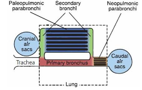
■ FIGURE 10-27 A schematic representation of the avian lung and air sacs. Color differences are for structural identification. The blackened areas correspond to blood capillaries and the adjacent blue areas correspond to the air capillaries. The combination of blood and air capillaries, compose the parabronchial mantle. The air sacs are extensions from the lungs, acting as bellows to create air flow. (Modified from Fedde MR. Respiration in birds. In: Swenson MJ, Reece WO, eds. Dukes’ Physiology of Domestic Animals. 11th edn. Ithaca, NY: Cornell University Press, 1993. Used by permission of the publisher, Cornell University Press.)
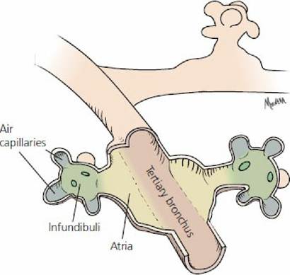
■ FIGURE 10-28 Schematic representation of tertiary bronchi and their extensions. A. Transverse section. B. Sagittal section. The atria are outpocketings from the tertiary bronchi. The infundibuli extend from the atria and have a number of extensions known as air capillaries. The air capillaries are interlaced with the blood capillaries (not shown).
The association of air capillaries with blood capillaries is known as the parabronchial mantle.
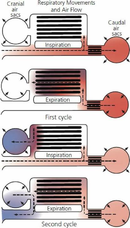
■ FIGURE 10-29 Pathway of air flow associated with inspiration and expiration in birds. The same bolus of air (colored area) (blackened area represents air capillaries) is followed through two respiratory cycles. It can be seen that ventilation of the parabronchial mantles is accomplished during inspiration and during expiration. Air going to the caudal air sacs ventilates the neopulmonic mantle, and as it leaves it ventilates both neopulmonic and paleopulmonic mantles. When the cranial air sacs expand during inspiration, they are filled by air that has passed through the parabronchial mantles. Cranial air sac air is then directed to the exterior during expiration without ventilating parabronchial mantles. (Modified from Scheid P, Slama H, Piiper J. Mechanisms of unidirectional flow in parabronchi of avian lungs: measurements in duck lung preparations. Respir Physiol. 1972; 14: 83-95.)
There are nine air sacs: two cervical, an unpaired clavicular, two cranial thoracic, two caudal thoracic, and two abdominal air sacs. The air sacs occupy space in the thoracic and abdominal cavities, and many have diverticula (extensions) into many of the bones replacing the bone marrow, causing them to be pneumatic. Replacing the bone marrow with air makes the bird lighter, possibly an assistance to flight. In the domestic species, the most prominent pneumatic bone is the humerus.
The air sacs are mucoserous sacs regarded as continuations of secondary bronchi beyond the lungs. Their walls are thin and have a poor blood supply. Because of their poor blood supply, air sacs are vulnerable to infection and to a condition known as air sacculitis. There is no significant gas exchange taking place in the air sacs. They do increase and decrease volume during respiratory cycles and, by this bellows-like action, function to increase pulmonary,ventilation.
Mechanics of Respiration and Air Circulation
1. Is diaphragm contraction a factor in avian respiration?
2. Describe how body volume changes influence inspiration and expiration.
3. Has air that enters the caudle and cranial air sacs been through a parabronchial
mantle?
4. Does air that leaves the caudle air sacs go through parabronchial mantles?
5. Does air that leaves the cranial air sacs go through parabronchial mantles?
6. Study Figure 10-30 to understand how blood capillary blood leaving the lung can have a lower Pco2 and a higher Po2 than gas that leaves the parabronchi.
7. How can blowing off excess Co2 during heat stress lower bicarbonate concentration (think hydration reaction)?
8. What is meant by a statement that notes the utilization coefficient for most birds is about one-half, versus one-fourth for mammals?
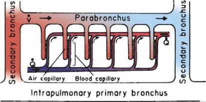
■ FIGURE 10-30 Schematic model of the cross-current gas exchange system in the avian lung. is the blood perfusion of the parabronchial mantle and represents the convective flow of gas through the parabronchus. Because of this arrangement with progressive incremental diffusion, blood leaving the parabronchial mantle has a higher Pao2 and a lower Paco2. (From Fedde MR. Respiration in birds. In: Swenson MJ, Reece WO, eds. Dukes’ Physiology of Domestic Animals. 11th edn. Ithaca, NY: Cornell University Press, 1993. Used by permission of the publisher, Cornell University Press.)
Birds have no diaphragm and therefore there is no separation between the abdominal and thoracic cavities. Accordingly, the entire body volume is changed during each respiratory cycle. The energy for the body volume change is derived from skeletal muscles in the body wall. Air flows through avian lungs during each expiratory phase and inspiratory phase of the respiratory cycle.
During expiration the body wall muscles contract, causing the body volume to decrease. The decrease in body volume increases air sac pressure, forcing the air within to flow back through the lungs and into the environment. Inspiration follows when the body wall muscles relax and body volume increases. Body volume increase is followed by a decrease in its pressure, which is followed by expansion of the air sacs and a decrease in their pressure. The decreased pressure allows air to flow through the lungs and into the air sacs.The passage of a single breath of air through the whole system of airways, air sacs, and lungs requires two respiratory cycles and is illustrated in Figure 10-29, where one bolus of air is followed through two respiratory cycles, from its entrance during inspiration of the first cycle to its exit during expiration of the second cycle. Notice that air entering the caudal air sacs during inspiration of the first cycle has been subjected to gas exchange via the neopulmonic parabronchial mantle and it is aerating the lungs via the neopulmonic and paleopulmonic mantles during expiration of the first cycle. In the second cycle, gas is received from the paleopulmonic mantle by expansion of the cranial air sacs during inspiration and evacuated to the environment by compression of the cranial air sacs during expiration.
Gas exchange between blood capillaries and air capillaries is illustrated in Figure 10-30. Air moves through the parabronchi by convection and into the air capillaries by diffusion. Blood in blood capillaries perfusing parabronchial mantles are partitioned into blood increments so that each increment of blood surrounds separate air capillaries throughout the length of the parabronchus. This arrangement, whereby air capillary gas flows through a parabronchus at right angles to the flow of blood, is known as cross-current flow. As gas flows through the parabronchi, CO2 is continuously diffusing from the blood capillaries, where the CO2 is higher, to the air capillaries, where the CO2 is lower, and O2 is continuously diffusing from the air capillaries, where the O2 is higher, to the blood capillaries, where the O2 is lower.
Because of this arrangement, the continuous loss of CO2 and gain of O2 causes the PaCO2 to be lower and the PaO2 to be higher when leaving the parabronchial mantle (see Figure 10-30). The cross-current arrangement is more efficient than gas exchanges in the mammalian lung and is most apparent when ventilation is increased in response to low oxygen (i.e., high altitude). Under these conditions blood capillary arterial PO2 may be only a few millimeters of mercury less than air capillary Po2 entering,the parabronchi. Oxygen will continue to be extracted as it proceeds through the parabronchial mantles regardless of the reduced pressure diffusion potential between blood capillary Po2 and air capillary Po2.General Considerations
• Mechanical valves to direct air flow have not been found in birds, and it is believed that the unidirectional flow of gas is probably governed by processes that create aerodynamic valves.
• Birds have a respiratory center and, similar to mammals, have chemoreceptors for CO2 and O2 that influence the response of the respiratory center.
• Unlike mammals, birds have CO2 receptors in their lungs that detect the CO2 levels in lung air. There is maximum receptor activity when CO2 is low, and this causes inhibition to respiration. It is believed that these receptors may serve to fine tune the pattern of respiration in birds.
• Diving ducks (not dabbling) have respiratory center sensitivity to postural changes (stretching of the neck, experimentally or naturally, as in diving, produces apnea).
• Ventilation of the lungs can be impaired by restricting movement of the sternum because the sternum must have a downward and forward movement to assist body volume increase needed during inspiration. This is an important consideration during bird restraint.
• Air sac infections can seriously impair ventilation, particularly if exudate plugs the entry from the air sacs to the lungs (common in aspergillosis, a fungus infection).
• Hyperventilation caused by heat stress reduces the Pco2 and bicarbonate concentration. The loss of bicarbonate causes egg shells to be thinner, and greater breakage occurs.
• High Paco2 causes cerebral vasodilatation and an increase in cerebral blood flow, while low Paco2 causes vasoconstriction and decreased cerebral blood flow. Mammals can tolerate a Paco2 down to 20 mm Hg, but below this the resistance to cerebral blood flow is so high that blood flow to the brain is compromised and results in cerebral ischemia. Birds are able to maintain cerebral blood flow even when Paco2 is 8 to 10 mm Hg. This allows birds to hyperventilate to meet oxygen demands while preserving cerebral perfusion, an obvious survival advantage for birds that fly at high altitudes.
• During the processing of poultry, birds are shackled, electrostunned, and exsanquinated, after which the carcasses are immersed in a scalding tank to assist with defeathering. At some part of the processing, there may be a variable amount of respiratory activity from agonal gasping, which results in aspiration of water with its contaminants with distribution to the air sacs. Although the air sacs are removed during evisceration, the diverticula that extend to the wing, thigh, and pectoral areas remain and become part of the edible tissue.
■