BLOOD SUPPLY
The vascular system of the udder is a complex network that ensures the delivery of nutrients and the removal of waste products, vital for the maintenance and production of milk. A modestly performing dairy cow is expected to generate at least 500 L of blood through its udder for each liter of milk.
This corresponds to around 10% of cardiac output during nursing. The caudal aorta provides the blood supply for the cow’s udder. It then splits into the right and left iliac arteries and flows bilaterally to supply the udder’s left and right parts. The iliac arteries bifurcate into external and internal iliac arteries. The perineal artery, a branch of the internal iliac artery, supplies blood to the caudal udder.The majority of blood supply to the mammary gland comes from the external iliac artery, which transforms into the prepubic artery until it passes through the internal inguinal ring, where it becomes the external pudendal artery and enters the mammary gland as the mammary artery. The external pudendal artery, branching from the pudendoepigastric trunk, plays a crucial role in supplying blood to the udder (Figure 22.1). Its division into cranial and caudal branches allows for distributed blood flow to both the anterior and posterior parts of the udder on the same side of the artery. The mammary artery further divides into cranial and caudal mammary arteries, which supply different areas of the udder. The right and left mammary arteries interconnect behind the medial suspensory ligament, and collateral circulation potential exists via the perineal artery connecting with the caudal mammary artery. Branches of the mammary arteries form a plexus above the teat, supplying the teat walls.
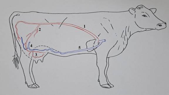
FIGURE 22.1 Vascular supply to and from the mammary Gland. 1-caudal artery; 2-external iliac & pudic artery; 3-cranial & caudal mammary artery; 4-external iliac & pudic vein; 5-subcutaneous abdominal vein.
The venous drainage from the udder is largely by way of a venous circle at the base of the udder, where it attaches to the abdominal wall. This venous circle is formed from the main veins that drain the udder. The external pudendal vein of each side receives blood from both the cranial and caudal quarters of the same side. Cranially, each external pudendal vein is continuous with the caudal superficial epigastric vein and caudally with the perineal vein. An anastomosis between the two caudal superficial epigastric veins just at or in front of the udder completes the venous circle.
The caudal superficial epigastric vein passes forward in a sagittal plane lateral to the midline on the ventral abdominal wall and joins with the cranial superficial epigastric vein, which ultimately drains to the internal thoracic veins and then to the cranial vena cava. Before the heifer comes into milk, the connection between cranial and caudal superficial epigastrics is poorly developed. During the first pregnancy, when the udder undergoes a marked increase in size and, consequently, blood supply, the two veins develop a functional anastomosis, after which they collectively constitute the subcutaneous abdominal vein or the milk vein. In high-producing dairy cows, sheep, and goats, the subcutaneous abdominal vein is large and tortuous. It passes through a foramen in the rectus abdominis muscle (the milk well), joins the internal thoracic vein, and ultimately drains into the cranial vena cava.
22.5