BODY CAVITIES
1. What are the subdivisions of the ventral body cavity?
2. Differentiate between visceral and parietal pleura.
3. What is the mediastinal space?
4. What structures occupy the mediastinal space?
5.
Differentiate between the abdominal and pelvic cavities with regard to the structures contained in each.6. What is the peritoneum?
7. Differentiate among omentum, mesentery, and ligaments.
A median plane view would show two main body cavities, the dorsal and ventral, and each has its subdivisions. The dorsal cavity contains the brain in its cranial cavity and the spinal cord in its vertebral cavity. The ventral cavity is subdivided by the diaphragm into the thoracic cavity cranially and the abdominal and pelvic cavities (collectively known as the abdominopelvic cavity) caudally.
Thoracic Cavity
The thoracic cavity is divided into two lateral chambers. Each chamber is lined by a serous membrane called the pleura and is termed a pleural cavity. The right and left lungs occupy their respective cavity and are enveloped by visceral pleura, which is continuous with the parietal pleura (mediastinal, costal, and diaphragmatic). The envelopment occurs during embryonic development. An analogy is that of pushing one’s fist into a partially inflated balloon, as shown for the heart in Figure 1-14. The space between the two lungs is known as the mediastinal space or mediastinum (Figure 1-15). It is a partition between the two pleural cavities. The heart, thoracic parts of the esophagus, trachea, vessels, and nerves are contained in the mediastinum, which is bounded laterally by mediastinal pleura. The mediastinal pleurae are the parietal pleurae that cover the sides of the partition between the two pleural cavities and the costal pleurae line the walls of the thorax. In general, the partition completely separates the right and left pleural cavities for all of the domestic animals except the dog and horse.
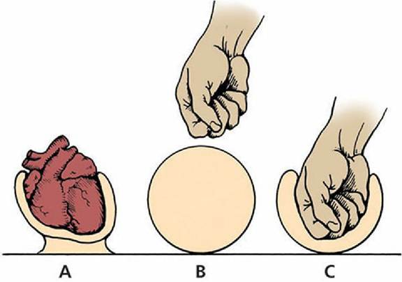
■ FIGURE 1-14 Invagination of the serous membrane to form outer (parietal) and inner (visceral) layers (A). Development proceeded similar to a fist being pushed into a balloon (B and C). (From Frandson RD, Wilke WL, Fails AD. Anatomy and Physiology of Farm Animals. 7th edn. Ames, IA: Wiley-Blackwell, 2009.)
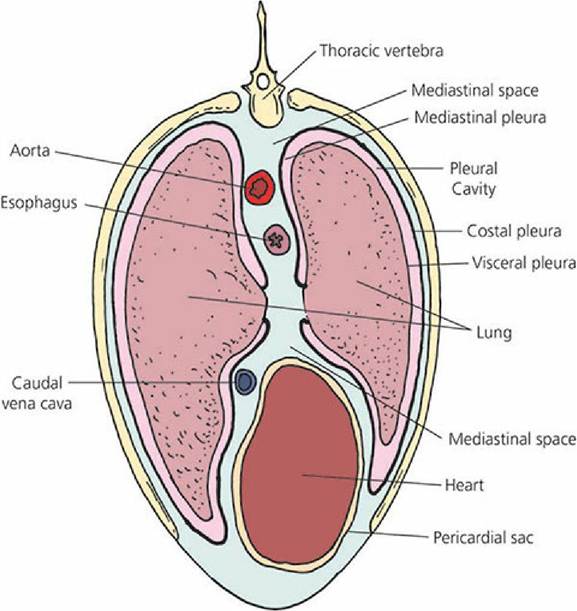
■ FIGURE 1-15 Schematic transverse plane of the equine thorax. The thoracic portions of esophagus, aorta, caudal venae cavae, and the heart are shown in the mediastinal space.
The Abdominopelvic Cavity
The abdominal cavity contains the kidneys, most of the digestive organs, and parts of the internal reproductive organs in both sexes. The pelvic cavity contains the rectum (terminal part of the gastrointestinal tract) and the internal parts of the urogenital system not otherwise found in the abdominal cavity. A serous membrane similar to that surrounding the heart and lungs is also found in the abdominopelvic cavity and is known as peritoneum.
The Peritoneum
The peritoneum lines the abdominal cavity and extends into the pelvic cavity. The abdominal organs begin development in a subserous (outside of the peritoneum) location, near the body wall. During development the organs enlarge and migrate into the abdominal cavity. They carry the peritoneum before them (introversion) and folds are formed that suspend them from the wall (Figure 1-16). The connecting folds are termed omenta, mesenteries, and ligaments. They contain a varying amount of connective tissue, fat, and lymph glands, and provide a pathway for vessels and nerves of the organs. An omentum is a fold that passes from the stomach to other viscera (soft structures). A mesentery is a fold that attaches the intestine to the dorsal wall of the abdominal cavity. Ligaments are folds that pass between viscera, other parts of the digestive tube, or connect them with the abdominal wall.
The coronary ligament (see Figure 1-16) is a sheet of peritoneum that passes between the diaphragm and the liver around the caudal vena cava.
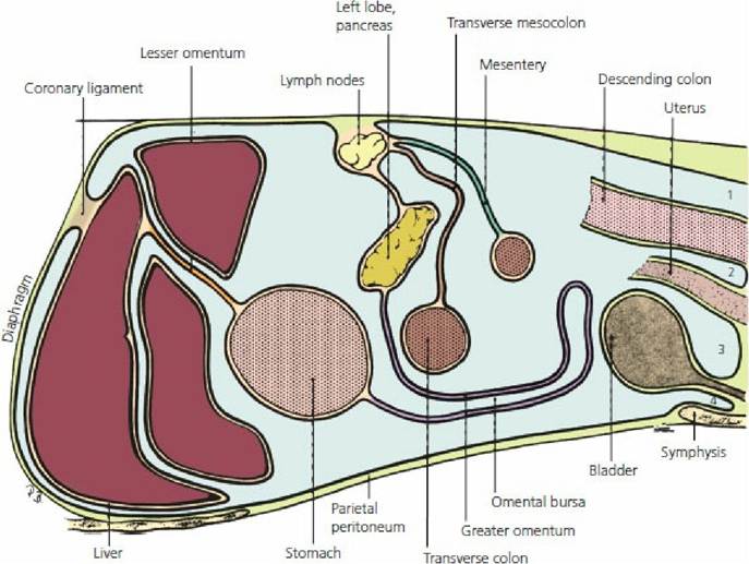
■ FIGURE 1-16 Schematic sagittal plane of the abdominal cavity showing the peritoneum and its connecting folds. (From Evans HE, deLahunta A. Guide to the Dissection of the Dog. 8th edn. St Louis, MO: Elsevier, 2017.) 1. Pararectal fossa. 2. Rectogenital pouch. 3. Vesicogenital pouch. 4. Pubovesical pouch.
■