CLINICAL ASSESSMENT OF CARDIOVASCULAR FUNCTION
The clinical assessment of cardiovascular function in animals, including dogs and cats, involves a combination of physical examination and diagnostic tests to evaluate heart structure, function, and overall cardiovascular health.
Here’s an overview of the clinical assessment methods used in veterinary medicine:4.10.1 Physical Examination
A. Inspection:
• Observation of general appearance, body condition, and demeanor.
• Assessment of respiratory rate, effort, and pattern.
• Evaluation of mucous membrane color (e.g., pale, cyanotic), capillary refill time, and presence of edema.
B. Palpation:
• Palpation of peripheral pulses (e.g., femoral, dorsal pedal).
• Assessment of heart rate, rhythm, and strength of cardiac contractions.
• Detection of abnormalities such as murmurs, thrills, or palpable masses.
C. Auscultation:
• Auscultation of heart sounds (S1, S2) using a stethoscope.
• Identification and characterization of additional heart sounds (e.g., S3, S4) or murmurs.
• Assessment of lung sounds (e.g., crackles, wheezes) and presence of murmurs transmitted to the lungs.
4.10.2 Diagnostic Tests
A. Electrocardiography (ECG):
• ECG records the electrical activity of the heart, providing information about heart rate, rhythm, and conduction abnormalities.
• Common ECG abnormalities include arrhythmias (e.g., sinus arrhythmia, atrial fibrillation), conduction delays, and chamber enlargement.
B. Thoracic Radiography (X-rays):
• Thoracic radiographs are used to evaluate heart size, shape, and position, as well as pulmonary vasculature and lung parenchyma.
• Findings may include cardiomegaly (enlarged heart), pulmonary edema, pleural effusion, or evidence of congenital heart disease.
C. Echocardiography (Cardiac Ultrasound):
• Echocardiography allows for non-invasive assessment of cardiac structure and function, including chamber dimensions, wall thickness, and valve morphology.
• Doppler imaging can evaluate blood flow velocity, gradient across valves, and presence of regurgitation or stenosis.
• Echocardiography is essential for diagnosing and monitoring congenital heart disease, acquired heart disease, and evaluating response to treatment.
D. Blood Pressure Measurement:
• Blood pressure measurement is important for assessing systemic perfusion and monitoring response to therapy.
• Techniques include Doppler ultrasonography, oscillometric devices, or direct arterial catheterization.
• Hypertension is a common finding in animals with chronic kidney disease, hyperthyroidism, or primary hypertension.
E. Blood Tests:
• Complete Blood Count (CBC) and Serum Biochemistry:
- CBC can detect anemia, leukocytosis, or thrombocytopenia, while serum biochemistry assesses renal and hepatic function, electrolytes, and biomarkers of heart disease.
• NT-proBNP (N-terminal pro-B-type natriuretic peptide):
- NT-proBNP is a biomarker released by the heart in response to myocardial stretch and stress and is used as a screening tool for heart disease and monitoring response to treatment.
F. Holter Monitoring:
• Holter monitoring involves continuous ECG recording over a 24-hour period, allowing for detection and characterization of arrhythmias or rhythm disturbances during normal daily activities.
G. Exercise Tolerance Testing:
• Exercise tolerance testing assesses cardiovascular function under controlled conditions, evaluating exercise capacity, heart rate response, and presence of exercise-induced arrhythmias.
H. Cardiac Catheterization and Angiography:
• Invasive procedures used for precise assessment of cardiac anatomy, hemodynamics, and
TABLE 4.2
Various Cardiovascular Diseases
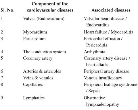
coronary artery blood flow. They are typically reserved for specialized centers and complex cases.
The combination of physical examination findings and diagnostic test results allows veterinarians to diagnose cardiovascular diseases, assess disease severity, and develop appropriate treatment plans tailored to the individual needs of the patient. Regular monitoring and follow-up evaluations are essential for managing chronic cardiovascular conditions and optimizing patient outcomes (Table 4.2).
4.11