Clinical Importance of the Ventral Body Wall
A surgical incision in the abdominal wall is called a laparotomy. It may be made in the midline, to either side of the midline or in the flank on either side. The choice of location of the laparotomy depends on a number of factors:
i) The avascularity of the linea alba resulting in slow healing; this is a particular problem especially in cattle where the linea alba is extensive.
ii) The bulk and weight of the abdominal contents leading to slow healing and risk of herniation.
iii) In the dog the ventral sheath of the rectus abdominis is particularly strong, and failure to suture this may result in breakdown of a midline incision.
iv) In a midline incision contraction of the muscles of the abdominal wall tends to retract the wound edges laterally.
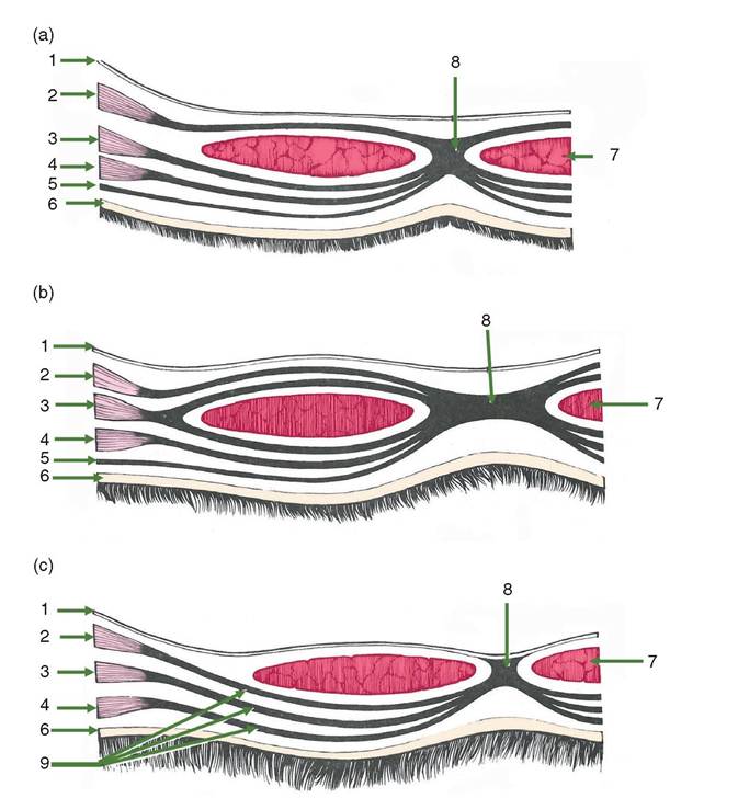
Figure 1.10 Transverse sections through the ventral body wall to show the species variation in the sheath of the rectus abdominis. (a) The horse, (b) the ox and (c) the dog (caudal third of abdomen only). 1 = parietal peritoneum; 2 = transverse abdominis muscle; 3 = interior oblique abdominis muscle;
4 = exterior oblique abdominis muscle; 5 = yellow abdominis tunic; 6 = skin; 7 = rectus abdominis muscle; 8 = linea alba; 9 = ventral sheath of rectus abdominis muscle
v) Flank incisions should be parallel to the muscle fibres to minimise bleeding from the vascular muscular tissue.
vi) In the cow a further complication of a midline incision is that branches of the mammary vein may cross the midline to anastomose with the opposite mammary vein.
1.6