Fractures and Fracture Healing
A fracture of bone is simply a break in the continuity of a bone. Among the many types of fractures described are the following (Fig. 5-6):
A simple fracture is one in which the skin over the fracture site is unbroken.
An open fracture is one in which a wound from the exterior contacts the bone at the point of the fracture. This may be caused by a broken end of bone perforating the skin or by a penetrating object, such as a bullet, causing the fracture.
A greenstick fracture is one in which one side of the bone is broken or splintered and the other side only bent. This type of fracture usually is found only in young animals.
A complete fracture is one in which the bone is broken entirely across.
A physeal fracture (formally known as epiphyseal fracture) is one that occurs at the junction of an epiphysis and the diaphysis of a bone. This type of fracture is limited to young animals.
A comminuted fracture is one in which the bone was splintered or crushed, producing small fragments.
if the broken ends of a fractured bone are brought into apposition (touch) and are immobilized (prevented from moving), the normal process of healing will take place (Fig. 5-7). when the fracture occurs, some blood vessels
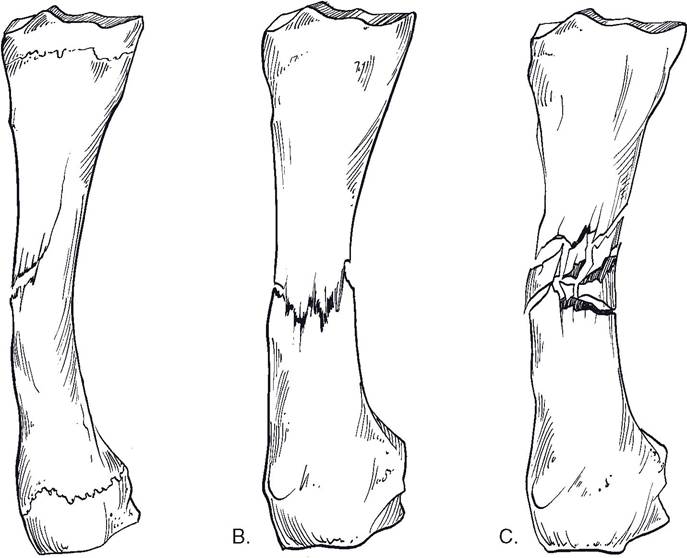
Figure 5-6. Types of fractures. A) Greenstick. B) Complete. C) Comminuted.
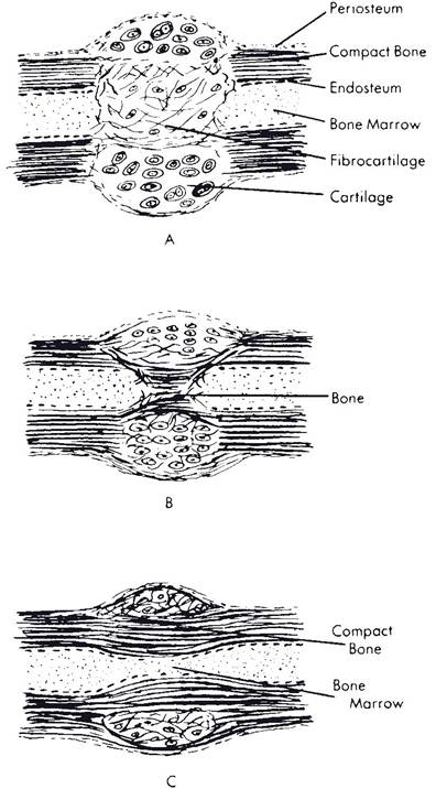
Figure 5-7. Some stages in healing of a fracture of a long bone. A) Early soft callus replaces blood clot. B) Intermediate callus. C) Nearly healed hard callus.
are ruptured, releasing blood around the broken ends of the bone. This forms a clot that is invaded by connective tissue cells forming granulation tissue (term for mass of tissue consisting largely of fibroblasts and capillaries).
The osteoblasts from the surface of the bone, from the periosteum, and from the endosteum lining the marrow cavities and haversian canals divide rapidly and produce a massive amount of osteoid tissue called a callus. The osteoid tissue fills the gap between the broken ends of the bone, fills the marrow cavity for a distance, and completely encircles the broken ends of the bone, forming an effective splint that usually prevents movement between the segments. The callus becomes mineralized, changing into true bone. Remodeling of the callus to form a typical bone shaft with a marrow cavity completes the healing process. Misalignment of the fractured bone is corrected to some extent by the action of osteocytes and osteoclasts, which also remove excessive internal and external callus. As soon as the bone is put to use, functional orientation of the callus begins, with a tendency to straighten imperfections in the alignment of the bone. The callus will increase in size on the concave side, where stress is greatest, and tend to erode on the convex side, thus tending to correct any deformity.The amount of spontaneous correction that is possible in fractures depends on a number of factors, including age of the animal, blood supply to the bone, degree of correction necessary, presence or absence of infection, and amount of damage to surrounding tissues. Excessive separation of fragments, which may be caused by too much traction or incomplete immobilization of a fracture, may result in nonunion, with fibrous tissue filling the gap between fragments.
Quickest fracture healing occurs in young animals, particularly if the fracture site has a good blood supply and is completely immobilized with the ends of the fragments in apposition. in humans, a fracture may heal completely within a month in an infant, but a similar fracture in a person past middle age may require 6 months or longer to heal.
if bone healing is delayed or a major defect has resulted from a severe fracture or surgical removal of bone, grafting or transplanting bone into the damaged area may stimulate healing. Bone for grafts may be obtained from the same animal that is receiving the graft (autogenous graft) or from another animal of the same species (allograft). Autogenous grafts are typically cancellous bone obtained from a site such as the proximal end of the humerus. Allografts of cortical bone may be used relatively intact or as bone chips to fill deficient areas. Autogenous grafts have an advantage in that the portion of the bone in contact with tissue fluid may survive and the osteoblasts become active. At the same time, osteoclasts remove the dead portions of the graft, which are replaced by healthy bone if the graft is functional and subjected to the proper amount of stress. Osteoblasts in allografts die, because the animal body tends to reject any foreign protein.