GENESIS OF MAMMARY gland (Mammogenesis)
Successful lactation is vital for the completion of the reproductive cycle in many mammalian species, with the processes of reproduction and lactation intricately linked. The journey begins with coordinated growth processes that commence in utero and extend through the delivery of the first offspring and into early lactation.
This growth and development of the mammary gland, known as mammogene- sis, lays the foundation for milk production. Lactogenesis marks the onset of lactation, during which alveolar cells undergo differentiation into milk-producing and -secreting cells. Galactopoiesis encompasses the maintenance and potential enhancement of established lactation. Following the cessation of milking, involution occurs, involving anatomical and physiological changes in the mammary gland, both immediate and long-term. These lactation processes, from mammogenesis to galactopoiesis and involution, are intricately regulated by hormonal interactions with the mammary gland and exchanges with other body tissues. Key endocrine glands involved in successful lactation include the hypothalamus, pituitary, adrenal glands, ovaries, and placenta, while other endocrine glands also exert influence.22.11.1 Pre-natal Development
The initial formation of the mammary band is observed, followed by the appearance of a streak within the band. The transformation of this streak into the mammary line is noted. After a certain stage, the line progresses into a mammary crest, which eventually develops into two or three layers. Subsequently, the mammary crest forms a hillock. The hillock then gives rise to a small bud, which emerges in the middle. A solid core of epithelial cells forms the primary sprout. The secondary sprout is characterized by the canalization of the teat area, as depicted in the figure. The teat pores are formed during the tertiary sprout stage.
The proliferation and eversion of epithelial cells demonstrate notable differentiation from the bud stage to the secondary sprouts, as illustrated in the Figure 22.4.22.11.2 Birth to Puberty
During the initial stage of development, from birth to puberty, young animals possess well-formed teats, although their glandular tissue remains rudimentary. Throughout the prepubertal period, which extends from birth until sexual maturity, mammary growth follows a pattern closely aligned with overall body growth. This phase is primarily characterized by the elongation of ducts and the maturation of stromal tissue, pivotal for future mammary function. Notably, the nutritional intake during this phase emerges as a critical determinant, as excessive feeding may lead to undesirable fat accumulation, potentially impeding the development of mammary tissue.
22.11.3 Puberty to Conception
As animals transition from puberty to conception, mammary growth undergoes a notable shift towards a more accelerated trajectory, marked by a rate surpassing overall body growth. This phase witnesses significant advancements in parenchymal growth, chiefly characterized by the elongation and branching of ducts, accompanied by concurrent stromal enhancement, particularly in blood vessel formation. Estrogen assumes a pivotal role during this period, fostering heightened mammary growth, particularly in females. The intricate interplay between estrogen and other regulatory hormones influences the branching patterns of mammary ducts, laying the groundwork for future milk production.
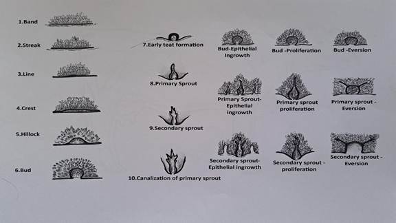
FIGURE 22.4 Diagram showing different stage of mammary gland development.
22.11.4 Conception to Parturition
The period spanning from conception to parturition signifies a crucial phase in mammary development, where factors associated with the maternal-conceptus axis exert profound influences on growth. This period witnesses exponential mammary expansion, with a substantial portion occurring during the latter stages of pregnancy.
Hormones such as estrogen, progesterone, prolactin, growth hormone, and placental lactogen orchestrate mammary development, facilitating the formation of extensive lobuloalveolar structures. Alveolar cells undergo proliferation and differentiation under the influence of these hormones, priming the mammary glands for milk secretion postpartum. The orchestrated hormonal profile, coupled with adequate nutritional support, is imperative for optimal mammary growth and subsequent lactation.22.11.5 Hormonal Influence of Mammary
Gland Development
Role of different hormones and their function in mammary gland is enlisted in Table 22.2. Follicle Stimulating Hormone (FSH) and Luteinizing Hormone (LH), produced by the adenohypophysis, regulate ovarian hormones such as estrogen and progesterone, influencing their effects on mammary gland development. Additionally, placental lactogen serves as a mammotrophic hormone.
The adenohypophysis exerts direct influence on the mammary gland through prolactin and somatotropic hormone, and indirectly through its control over thyroid and adrenal cortex hormones.
22.11.6 Growth Factors of Mammary
Gland Development
Various unidentified growth factors have been proposed for both normal and cancerous mammary cells based on activities observed in tissue extracts and conditioned medium. These factors originate from non-mammary tissues, such as the pituitary and liver (endocrine), as well as from the mammary gland itself (paracrine or autocrine), associated with mammary mesenchyme or epithelium respectively. Until these factors are fully characterized, whether they function as cell nutrients or polypeptide growth factors, activating DNA synthesis in dormant cells via high-affinity receptors, remains unclear. Examples of tissue-derived mitogens identified as cell nutrients include phospho-ethanolamine from bovine pituitary and transferrin from pig pituitaries. Studies have demonstrated proliferation and improved epithelial morphology in mouse mammary epithelium cultures grown in or on collagen when exposed to adipose tissue- conditioned medium, suggesting a potential role for unsaturated fatty acids.
In addition, Insulin-like Growth Factors (IGF) and Epidermal Growth Factor (EGF) play significant roles..22.11.7 Fat Pad and Mammary Gland Significance
The fat pad in the mammary gland serves several important functions in mammary physiology and development:
• Structural Support: The fat pad provides structural support to the mammary gland. It helps maintain the shape and integrity of the glandular tissue, ensuring proper positioning and function of the mammary epithelial cells.
TABLE 22.2
Function of Different Endocrine Secretions in Mammary Gland
Endocrine component (organ)
FSH (Anterior pituitary)
Prolactin (Anterior pituitary)
GH (Anterior pituitary)
Oxytocin (Posterior pituitary)
T3, T4 (Thyroid)
Calcitonin (Thyroid)
Glucocorticoids (Adrenal cortex)
Epinephrine, norepinephrine (Adrenal medulla)
Estradiol (ovary)
Progesterone (ovary)
Estradiol (Placenta)
Progesterone (Placenta)
Placental lactogen (Placenta)
Modified and inspired from Singh and Roy (2017).
Function in mammary gland
Estrogen secretion from ovarian follicles
Mammary growth; initiation and maintenance of lactation
Stimulates milk production
Milk ejection
Stimulates O2 consumption, protein synthesis and milk yield
Calcium and phosphorus metabolism
Initiation and maintenance of lactation
Inhibition of milk ejection
Mammary duct growth
Mammary alveolar - lobule growth, inhibition of lactogenesis
Mammary duct growth
Mammary alveolar - lobule growth, inhibition of lactogenesis
Mammary growth
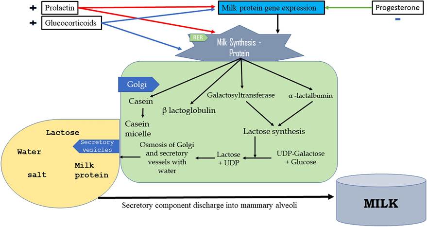
FIGURE 22.5 Flow diagram showing the overview of the lactogenesis process.
• Energy Storage: Adipose tissue within the fat pad serves as a reservoir for energy storage. During lactation, when there is a high demand for energy to support milk production, adipocytes in the fat pad can release stored triglycerides as fatty acids to provide energy for mammary epithelial cells.
• Insulation: The fat pad provides thermal insulation to the mammary gland, helping to maintain a stable internal environment. This is particularly important during lactation when the mammary gland is actively producing and secreting milk.
• Hormonal Regulation: Adipose tissue is an endocrine organ that secretes hormones and adi- pokines, which can influence mammary gland development and function. For example, adipose- derived hormones such as leptin and adiponectin may regulate aspects of mammary gland physiology, including lactation.
• Mechanical Protection: The fat pad can also provide mechanical protection to the mammary gland, cushioning it against physical trauma or injury.
Overall, the fat pad plays a crucial role in supporting mammary gland function and ensuring optimal lactation and reproductive success in mammals.
22.12