HEMATOPOIESIS (FORMATION OF BLOOD)
Few lymphocytes, out of all the cellular constituents, live for many years; all other cells have lifespans of hours to weeks and should be periodically replaced by newer cells. Hemopoiesis, also known as hematopoiesis, is the process that creates these cells and continues for the duration of an animal’s life.
The foetus’s liver and spleen are where hematopoiesis takes place. The majority of an infant animal’s body’s bones contain red bone marrow, which is where this process mostly takes place. Many bones are impacted by the increased demand for blood cells that occurs during growth and development. The rate of production falls because the animal isn’t growing as quickly as it is maturing. The highly vascularized connective tissue known as red bone marrow is found in the microscopic gaps between the trabeculae of spongy bone tissue. It is primarily found in the proximal epiphyses of the humerus and femur, pectoral and pelvic girdles, and bones of the axial skeleton. Hemocytoblasts, also known as pluripotent stem cells, make up about 0.05-0.1% of red bone marrow cells. They are derived from mesenchyme, the tissue from which practically all connective tissues develop. These cells have the ability to differentiate into a wide variety of cell types.When certain red bone marrow becomes less active, yellow bone marrow - which is primarily made up of fat cells - is produced. However, increased physiological demands can cause yellow marrow to reactivate. The type of bone marrow is so named because of how ugly it looks. Red bone marrow is found in different places in the adult bodies of different species. In most species, the red marrow sites are the skull, ribs, sternum, vertebral column, pelvis, and the proximal ends of the femurs.
Red bone marrow stem cells self-replicate, multiply, and differentiate into cells that give rise to mast cells, reticular cells, adipocytes, blood cells, and macrophages.
Certain stem cells have the capacity to differentiate into osteoblasts, chondroblasts, and muscle cells. These cells may find application in the replacement of bone, cartilage, and muscle tissue. The stroma, or framework, that holds up the red bone marrow cells is made of reticular fibers, which are produced by the reticular cells.Red bone marrow’s pluripotent stem cells divide into two additional types of stem cells, each of which has the potential to differentiate into a variety of cell types, in order to produce blood cells. These stem cells are referred to as lymphoid and myeloid stem cells. Red bone marrow is where myeloid stem cells start to develop into red blood cells, platelets, neutrophils, eosinophils, basophils, and monocytes. Red bone marrow is the starting point for lymphoid stem cell development, which is finished in lymphatic tissues where it gives rise to lymphocytes. Despite having unique cell identity markers in their plasma membranes, the different types of stem cells are not histologically distinct and instead resemble lymphocytes. These myeloid stem cells differentiate into precursor cells and progenitor cells
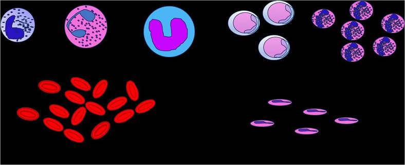
FIGURE 6.1 Diagrammatic presentation of formed components of blood. 1. White Blood Cells: Basophil (A), Eosinophil (B), Monocyte (C), Lymphocytes (D), Neutrophils (E). 2. Red Blood Cells. 3. Platelets
TABLE 6.1
Table Representing RBC Count in Different Species
| Species | RBC (millions) or (volume mm3) |
| Cattle, Pig, Dog, Cat | 6-8 |
| Sheep and Goat | 10-14 |
| Horse | 7-12 |
| Rabbit | 5.5-6.5 |
| Chicken | 2.5-3.2 |
during hemopoiesis. The progenitor cells, which are dividing into CFU-E (erythrocyte), CFU-Meg (megakaryocyte, or platelets), and CFU-GM (granulocyte), are what form the colonies. Monocytes and eosinophils develop as a result of the precursor cells dividing and forming blasts.
Various hormones play important roles in all these mechanisms, e.g., hemopoietic growth factor, erythropoietin, thrombopoietin, cytokines, interleukins etc.
6.8