Hypothalamopituitary Axis
The hypothalamus is ventral to the thalamus in the diencephalon and forms the floor and part of the wall of the third ventricle (see Chapter 9). The pituitary gland, or hypophysis cerebri, is attached to its base by the infundibulum, a stalk of nervous tissue (primarily axons).
Cell bodies of neurons whose axons form the infundibulum are found in the hypothalamus, and their termini abut on the capillaries in the neural part of the pituitary gland (neurohypophysis, posterior pituitary, or pars nervosa). Associated with the infundibulum is a unique system of arterioles and capillaries called the hypothalamohypophysial portal system. This system is a true vascular portal system in that blood from a capillary network in the hypothalamus flows through portal vessels (similar to veins) to the glandular portion of the pituitary (adenohypophysis, anterior pituitary, or pars distalis), where it enters a second capillary network (Fig. 12-3). During development, the pituitary gland forms by the conjoining of a ventral outpouching of the diencephalon (the future infundibulum and neurohypophysis) and a diverticulum, Rathke’s pouch, which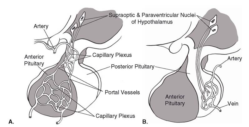
Figure 12-3. Relation between hypothalamus, neurohypophysis, and adenohypophysis. A) Drawing illustrates network of portal vessels. B) Drawing illustrates direct neural connection between hypothalamus and neurohypophysis.
arises from the nearby dorsal ectoderm of the pharynx. The cells of Rathke’s pouch become the adenohypophysis.
The neurotransmitters released by hypothalamic neurons whose termini end in the neurohypophysis enter the blood and are carried to distant sites to function as systemic hormones (Fig. 12-3). These peptides are manufactured by the neuronal cell bodies within the hypothalamus, transported via axons into the neurohypophysis, and released directly into blood vessels when action potentials arrive at the telodendria.
other hypothalamic neurons release neurotransmitters that are carried from the hypothalamus to the adenohypophysis via the hypothalamohypophysial portal system (Fig. 12-3). There, these neurotransmitters act on endocrine cells either to stimulate or to inhibit the release of other hormones (Table 12-1). While these neurotransmitters do not travel to a distant site to stimulate target cells, they are transmitted via the blood from their site of origin to their site of action. Thus, these neurotransmitters may be considered hormones, but they are also referred to as inhibiting or releasing factors. All of these neurotransmitter hormones except dopamine are small peptides, and the small peptides are rapidly degraded after passing through the hypothalamohy- pophysial portal system and entering the general circulation. The unique portal system permits the delivery of a relatively high concentration of the releasing or inhibiting factors to the adenohypophysis so that a biologic effect is possible.
The different cell types of the adenohypophysis exhibit different histologic staining characteristics, depending on the hormone they produce. As a consequence, cells are characterized as basophils, acidophils, or chromophobes, among other specific types. These endocrine cells are arranged in cords or clusters around blood-filled sinusoids, in keeping with their role as an endocrine organ. The neurohypophysis has the typical microscopic appearance of nervous tissue, consisting of unmyelinated axons and supportive glial cells.
Historically, the pituitary gland was known as the master gland because of the large number of hormones secreted and their wide-ranging effects. several hormones from the adenohypophysis stimulate distant endocrine glands to increase production of their own hormones (Table 12-1). These stimulatory adenohypoph- yseal hormones are often called trophic or tropic hormones.
The hypothalamus functions as a crucial interface between the nervous and endocrine systems, where sensory information is integrated and used to regulate the endocrine output of the pituitary gland.
Much of this information is related the status of the internal environment in question (e.g., extracellular fluid osmolality, blood glucose concentration, body temperature and metabolic rate). Release of adenohypophyseal hormones can also be regulated through more direct negative feedback loops based on the blood concentrations of the hormones involved. The hormone produced by the target endocrine gland of a specific trophic hormone can act on (1) the hypothalamus to reduce the production of its releasing factors and (2) the adenohypophysis to reduce its release of the tropic hormone (Fig. 12-4). The tropic hormones of the adeno-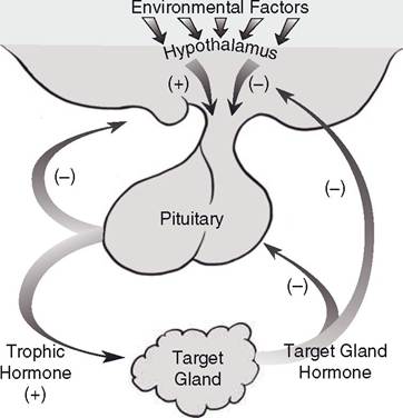
Figure 12-4. Potential feedback loops to regulate hypothalamic releasing hormones and tropic hormones from adenohypophysis. stimulation of secretion indicated by (+) and inhibition indicated by (-).
hypophysis may also reduce hypothalamic releasing factors via a short negative feedback loop.
A synopsis of the hormones released into the general circulation by the hypophysis, the factors that regulate their release, and their general functions is presented here. Further details on these functions will be given in subsequent chapters when the function of their target organs is covered. All of these hormones are peptide or protein hormones based on their chemical structure.