INTEGUMENT
The integument of the amphibian is arguably one of the most important organ systems. The skin functions not only in a protective capacity but also as a sensory organ, and plays vital roles in thermoregulatory and hydrational homeostasis, sex recognition, and reproduction.
Heatwole and Barthalamus (1994) provide an excellent review of the amphibian integument.Like that of all vertebrates, the amphibian's skin consists of an epidermal layer and a dermal layer. Although the epidermis consists of several cell layers, it is considerably thinner than that of other tetrapods, with the stratum corneum usually consisting of only a single layer of keratinized cells in most species. In fact, some aquatic salamanders lack keratinization of the stratum corneum altogether. Shedding of the stratum corneum occurs regularly, and most amphibians will eat their skin sheds (Goin et al. 1978; Weldon et al. 1993). The basal epithelium is four to eight cell layers thick, and is the site of epidermal regeneration. Although the epidermis provides some protection from abrasive substrates, the epidermis is easily damaged if the amphibian is improperly handled or is in contact with inappropriate substrates. The resulting damages from even an apparently minor injury can have serious consequences as there is no longer an effective barrier against opportunistic microorganisms.
The well-vascularized dermis consists of an outer spongy layer (the stratum spongiosum), and a more compact inner layer (the stratum compactum). Capillaries, nerves and smooth muscle are found throughout the dermis. Some caecilians possess tiny dermal scales, not found in the other two orders. Three types of chromatophores that are responsible for skin coloration, as well as specialized glands, are present in the stratum spongiosum. In caecilians and salamanders, the stratum compactum contains collagen fibers that tightly adhere it to the underlying connective tissue, musculature, and bones, while in anurans there is not a tight association, resulting in a potential subcutaneous space for fluid administration.
Due to this loose association, anurans (but not caecilians or salamanders) can appear edematous,CLINICAL NOTE
When handling amphibians there is always some damage done to the epithelium; therefore, it is recommended that lightly moistened, powder-free latex or nitrile gloves be worn to minimize damage to the sensitive skin and decrease the transfer of microorganisms, or potentially noxious substances, from the hands of the clinician (Fig. 1.12).
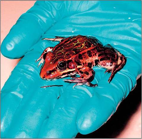
Figure 1.12 • Northern leopard frog (Rana pipiens) being appropriately handled with gloves. (Photo by Whiteside.)
CLINICAL NOTE
Amphibians are exquisitively sensitive to many toxic compounds at levels much lower than those that would cause clinical effects in higher vertebrates. It is also important to note that, owing to the thin nature of the epithelium, the skin represents an effective route for treatment in most amphibians, allowing topical administration of anesthetics such as MS-222 (tricaine methane sulfonate) or antibiotic, with resulting systemic effects (Whitaker et al. 1999; Whitaker & Wright 2001; Wright 1996, 2001c).
either as a result of normal water storage or due to pathological processes (Goin et al. 1978; Mitchell et al. 1988; Wright 1996, 2001c).
A variety of specialized glands are found within the epidermis and dermis. Some glands produce mucous or waxy substances to reduce evaporative water loss, as previously described. The dermis also contains numerous glands that produce toxic or irritating substances as protective mechanisms. Many of the glandular secretions of caecilians, salamanders, and anurans can be irritating to the mucous membranes of humans, while other amphibians, such as the arrow poison frogs (Dendrobates and Phyllobates spp.), produce steroidal alkaloid toxins that are potentially lethal to people. Some species, such as the fire salamander (Salamandra salamandra), can actually spray poison from dorsal glands, whereas others, such as the giant toad (Bufo marinus), have large parotid glands on the back of the neck that may spurt several feet when pressure is applied.
CLINICAL NOTE
The irritating, or even highly toxic, secretions from some amphibians are another reason why latex or nitrile gloves should be worn by the clinician, and in some cases eye protection also may be prudent (Goin et al. 1978; Mitchell et al. 1988; Whitaker et al. 1999; Wright 1996, 2001a, 2001c).
Amphibians
True scales and claws are lacking in amphibians, although some species have modified cornified epidermal claw-like structures, as seen in the African clawed frog (Xenopus laevis) and some salamanders, such as Onchydactylus spp. Other amphibians have different modifications, such as the cornified areas on the feet of Spadefoot toads (Scaphiopus spp. and Pelobates spp.) (Goin et al. 1978; Mitchell et al. 1988; Wright 1996, 2001c).
13
KEY POINTS
• The class name refers to the dual life stages: aquatic and terrestrial.
• Amphibians have a three-chambered heart (two atria and a ventricle).
• Aquatic larval forms use external gills for respiration and tend to be herbivorous, whereas terrestrial adults develop internal lungs and are carnivorous.
• All are poikilotherms (ectotherms).
• Gloves should be worn when handling amphibians to prevent damage to the patient and the handler.
• A specialized sensory organ in the oral cavity (Bidder's organ) is responsible for chemodetection.
• Most larval forms and some adult forms retain the ability to regenerate amputated tails, digits, and limbs.
• Phlebotomy sites include the heart, the ventral abdominal vein, the femoral vein, the lingual plexus, and the ventral tail vein (when present).