INTERNAL ANATOMY
The tissue that makes up the udder is pink and white. White tissues, or non-parenchymal cells, are composed of connective, fibrous, and fatty tissue, whereas pink tissue is known as parenchyma or epithelial tissue.
The parts of the udder’s internal structure are as follows:Teat Pore: During milking, the milk is drained out of the teat through this small orifice. The muscle known as the teat sphincter keeps it firmly covered. Between milkings, the muscles of the teat sphincter prevent milk from leaking.
The teat contains a portion of the canal called the “fat streak” that opens up in the teat cistern. There are unique folds at the intersection of the teat cistern and teat canal that stop milk flow against gravity. These folds are called Furstenberg’s Rosettes. One of the most significant defenses against microorganisms entering the udder is the teat canal. The teat canal ranges from 8 to 12 mm in length, and depending on the teat’s size, it is capable of holding 15 to 40 ml of milk. Slow milkers are cows with narrow teat canals or tight sphincter muscles, which make them take longer to milk. Large teat canals or weak sphincter muscle animals leak milk between milkings and are more susceptible to pathogens that cause mastitis. Additionally, the smooth muscles in the milk ducts and the vascular system are related to the bladder by nerves. Every teat has its own cistern for retaining milk, holding around 400-500 ml between milkings.
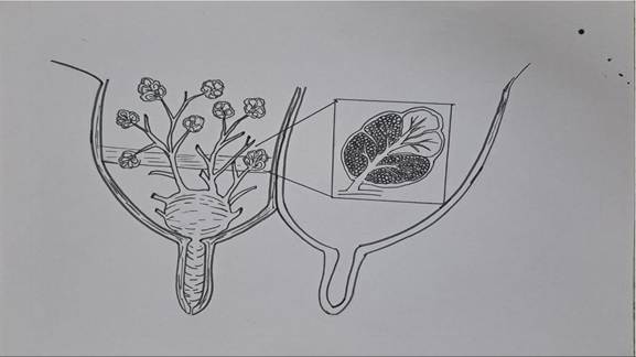
FIGURE 22.3 Diagram showing the internal structure of the mammary gland.
The gland cistern is partly divided by annular folds of tissue and is situated directly above the teat cistern. Milk is carried into the gland cistern via the giant milk ducts, which are opened by the small ducts. Each alveolus produces milk, which is transported by the tiny ducts.
The udder’s ductal system is made up of several varying-sized ducts arranged in the following order: lobar ducts, intralo- bar duct, interlobular duct, and intralobular ducts. The main function of the duct system is to collect milk from the secretory tissue, store a part of the milk between milkings, and transport the milk into the gland cistern. Constrictions are placed at the tip of both large and tiny ducts to stop milk from dripping and leaking into the teat.Lobes and Lobules: Each mammary gland comprises several lobes, and each lobe consists of numerous lobules. Layers of connective tissue divide lobes and lobules. A lobule can contain between 200 and 250 alveoli. Each alveolus is composed of a single layer of epithelial cells, responsible for producing milk, which is then discharged into the alveolar lumen.
The spherical alveolus of the udder is the functional unit responsible for milk production. It is lined by a single layer of epithelial cells, and a small duct called the alveolar duct drains the milk.
The outermost layer of the alveolus also contains muscle-like cells called myoepithelial cells, which contain receptors that bind to the hormone oxytocin, necessary for milk secretion. When oxytocin binds to these receptors, the myoepithelial cells contract, releasing milk into the alveolar lumen. The contraction of myoepithelial cells can increase intramural pressure to 60 mmHg or more, practically stopping milk secretion. Each quarter’s primary lobe is formed by a lobule, which consists of several alveoli. There are approximately 5 x 10^12 secretory cells in the udder’s alveolar tissue of dairy cows.
The mammary gland of a cow or buffalo is composed of two components: connective tissue, which is non-parenchy- mal, and pink secretory parenchymal tissue. The amount of milk produced by animals is determined by the number of secretory tissues or secreting cells. Thus, the rate-limiting component in the udder’s ability to produce milk is parenchyma. Contrary to popular assumption, a large udder does not necessarily indicate a great ability for milk production. There might not be as much space to accommodate milk-secreting tissue in an udder with a large volume of connective and adipose tissue. After milking, some udders shrink significantly and become flabby due to having less connective tissue. Milk is generated by the secretory cells, sometimes referred to as epithelial cells, which are organized in a spherical shape known as alveoli, forming a single layer atop a basal membrane.
22.9