INTERNAL ANATOMY OF KIDNEYS
The medial aspect of each kidney has a concavity, the hilus, where arteries and nerves enter the kidney, and the ureter, veins, and lymphatic vessels leave. The wide origin of the ureter in the kidney is the renal pelvis, which receives urine from the collecting tubules.
The cavity-like structure in the kidney that contains the pelvis is the renal sinus. The bovine kidney does not have a renal pelvis, so the ureter arises directly from the coalescence of individual calyces. The portion of the kidney immediately surrounding the renal pelvis is the renal medulla, which appears striated because of the radially arranged collecting tubules. The medulla also contains some loops of Henle (descending and ascending loops), and it is surrounded peripherally by the renal cortex in which the renal corpuscles are present (Figure 18.2).The cortex has a granular appearance because of the large number of renal corpuscles, as well as proximal and distal convoluted tubules and other segments of loops of Henle. The medulla and cortex are arranged in units called lobes, which is a cone-shaped aggregates of renal tissue. The medullary portion of each lobe constitutes a renal pyramid, whose apex, i.e., renal papilla, is directed toward the renal pelvis. In the bovine kidney, each pyramid is associated with one of the grossly obvious lobes, each of which communicates with a minor calyx. The renal cortex of the equine, ovine, and caprine kidneys lacks visible divisions from outside into individual lobes, so the kidneys in these species appear smooth. In these species, the tips of the renal
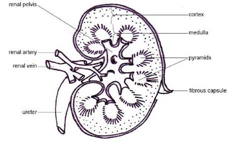
FIGURE 18.2 Internal structure of kidney
pyramids fuse with each other to form a common renal papilla, called the renal crest. The porcine kidney lacks external divisions into lobes, but the renal papillae of the medulla distinguish each lobe internally. In the kidney of the ox and pig, individual pyramids project into minor calyces (cup-like diverticula of the common collecting space within the renal hilus). These, in turn, empty into major calyces in these species empty directly into the ureter.
18.8