Key points
■ Twelve pairs of cranial nerves innervate the head and extend into the body.
■ Individual nerves have specific sensory and/or motor, somatic and/or autonomic functions.
■ Knowledge of the location and action of individual cranial nerves is critical for the interpretation of the neurological examination (Tables 10.1, 10.2).
■ Cranial nerves are numbered sequentially from rostral to caudal based on the site of their attachment to the brain. CNN I and II attach to the forebrain; CNN III and IV are associated with the midbrain (rostral brainstem); CN V to XII are associated with the mid and caudal brainstem.
■ Cranial nerves III-XII neurons are arranged in nuclei in the brainstem.
■ Cranial nerves and their CNS components are bilaterally paired.
■ Most cranial nerves, excluding the optic and olfactory nerves, have a peripheral portion that is ensheathed/myelinated by Schwann cells.
Table 10.1 Cranial nerves and their function
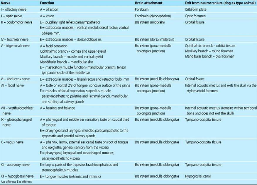
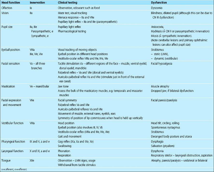
Table 10.2 Cranial nerve testing and sign of dysfunction
The vast majority of vertebrates have 12 pairs of cranial nerves (CNN), but there may be only ten pairs in amphibians and fish. Cranial nerves are numbered, using Roman numerals, for their point of attachment to the brain. The first cranial nerve (CN I) attaches to the most rostral aspect of the forebrain as the olfactory bulb, whilst CN XII is attached to the caudal medulla oblongata (Fig. 10.1). The others attach, in sequence, between these two end points.
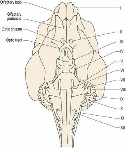
Fig. 10.1 Ventral aspect of canine brain and cranial cervical spinal cord depicting the attachment of the cranial nerves.
Named cranial nerve nuclei III-XII are found in the brainstem; they are bilaterally paired (Fig. 10.2). Their location relative to the sulcus limitans determines their function. Sensory cranial nerve nuclei are located dorsal to the sulcus limitans, parasympathetic (autonomic) nuclei are located lateral to it, and motor nuclei are ventral to it (Figs. 1.7, 2.3A) (Note: an exception to this is the location of the vestibular nuclei, which are sited dorsal and lateral to the sulcus limitans, despite having both sensory and motor functions). Remembering this anatomical arrangement is useful, as when the clinician is looking at a cross-section of the brainstem (e.g. MR image or tissue section), they can make an educated assumption about function of grey matter based on its dorsoventral position.
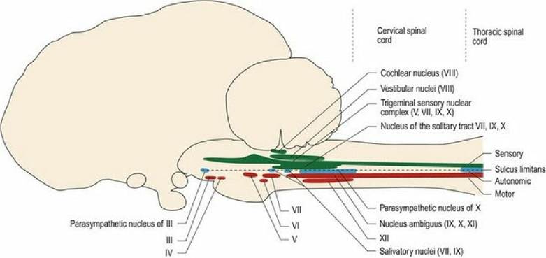
Fig. 10.2 The functional columns of grey matter in the spinal cord and their fragmentation to form nuclei
of the cranial nerves in the brain stem.