Lymphatic System
The lymphatic system includes the lymphoid tissues (e.g., lymph nodes and nodules and the spleen) and the lymphatic vessels distributed throughout the body (Fig. 16-2). It drains tissue fluid (called lymph within the lymphatic system) and is a framework for the circulation, production, and maturation of immune cells.
The tissue fluid-draining function of the lymphatic system augments the venous circulation and therefore assists in the control of interstitial fluid pressures. The lymphatic system is also an important component of immunologic defense of the body, as movement of lymph brings microorganisms and other foreign substances into contact with immune cells.Lymphoid tissue consists of accumulations of lymphocytes trapped in the spaces between fibers of reticular connective tissue. The lymphoid tissue may be scattered diffusely in some regions (as is commonly found in mucous membranes), may appear as nodules (as in the intestinal submucosa), or may be encapsulated to form specific organs, including lymph nodes, the spleen, thymus, and tonsils. The lymphatic vessels and tissues are arranged so that tissue fluid is exposed to aggregates of immune cells, which scrutinize the fluid for foreign cells and substances, thereby assisting in the control of infection.
Lymphatic Vessels
The lymphatic vessels constitute a one-way pathway that parallels the venous system and eventually empties into the cranial vena cava or some of its tributaries. The smallest lymphatics begin blindly between tissue cells as lymphatic capillaries, which collect the tissue fluid not absorbed by the venous system. When the tissue fluid enters the lymphatic vessels, it is known as lymph, which consists of fluid originally derived from the blood, a variety of blood cells, and sometimes microbes. Lymphatic vessels carry lymph back to the great veins of the heart.
Lymph Nodes
Lymph nodes are discrete knots of lymphoid tissue scattered along the course of lymph vessels. Lymph nodes filter the lymph and act as one of the first defenses against infection by harboring lymphocytes, plasma cells, and macrophages.
Each lymph node is surrounded by a connective tissue capsule that sends numerous connective tissue trabeculae into the substance of the node (Fig. 16-3). The node is roughly divided into cortex, paracortex, and medulla, with large numbers of lymphocytes and macrophages in all three. Lymphocytes in the cortex are arranged in nodules (Fig. 16-3). Dark-staining groups are primary nodules, and light-staining groups are secondary nodules. secondary nodules are areas of rapid B cell proliferation, and for that reason their cores are called germinal centers. The paracortex, deep to the cortex, is populated primarily with T lymphocytes and dendritic cells.
Lymphocytes in the medullary portion of the lymph node are arranged in medullary cords rather than nodules (Fig. 16-3). These tend to be primarily accumulations of plasma cells.
immediately deep to the capsule of the node is a space, the subcapsular sinus, which communicates with other sinuses of the cortex and medulla. Lymph delivered by afferent lymph vessels enters the subcapsular sinus and is slowly filtered through the cortex and medulla, to emerge finally at the hilus of the node, where blood vessels and nerves enter and the efferent lymphatic vessels emerge, carrying the lymph that has percolated through the node. This arrangement is ideally suited for the presentation to immune cells of antigens collected in the tissue fluid.
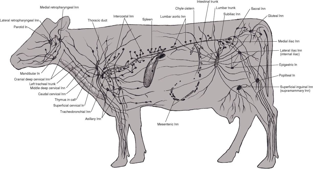
Figure 16-2. The lymphatic system of the cow. (Reprinted with permission of Wiley-Blackwell from McCracken, T.O., Kainer, R.A., and Spurgeon, T.S. Spurgeon’s ColorAtlas OfLargeAnimalAnatomy. Baltimore: Lippincott Williams & Wilkins, 1999.)
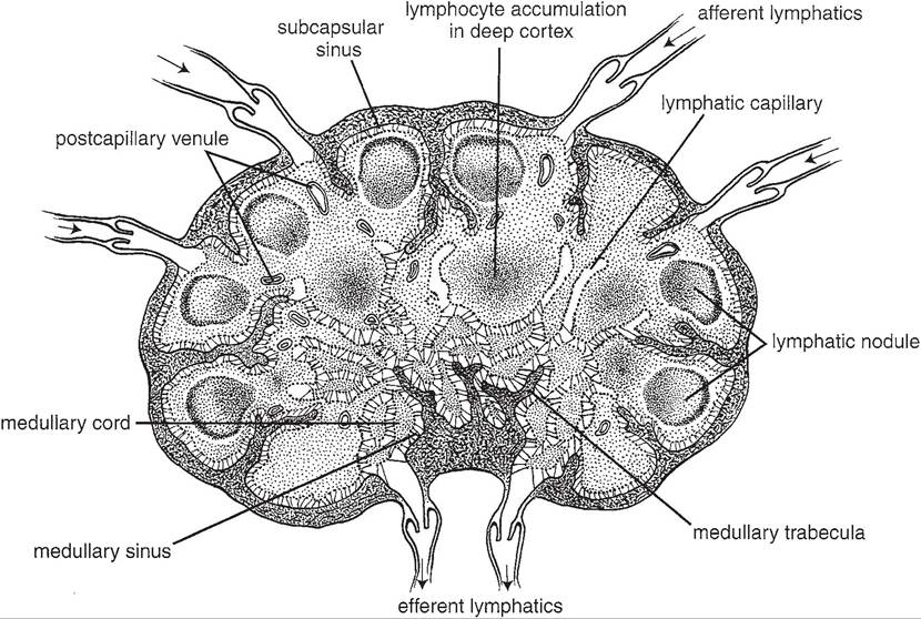
Figure 16-3.
Anatomy of a typical lymph node. (Reprinted with permission of Wiley-Blackwell from Eurell, J.A. and Frappier, B.L. DellmannS Textbook of Veterinary Histology, 6th ed. Ames, IA: Blackwell Publishing Professional, 2006.)Oddly enough, the porcine lymph node’s histologic architecture is a reverse of that seen in other species, with the nodules found in central regions and medullary cords on the periphery of the node. Flow of lymph likewise is reversed, with afferents entering at the hilus and percolating through the lymphatic tissue to emerge at the capsule.
Lymph nodes are scattered throughout the body, and in general their number and location are fairly consistent within a given species (Table 16-2). For convenience, groups of related nodes are often called a Iymphocenter. The condition of a lymph node often reflects health of the area from which it receives lymph. If a specific area is infected, the lymph nodes in that area tend to enlarge as the germinal centers begin producing additional lymphocytes in response to the antigens delivered to the node. For example, a horse with strangles, a bacterial infection of the nasal cavity and pharynx, frequently shows enlargement of the mandibular and retropharyngeal lymphocenters. The lymph nodes at these locations receive their afferent vessels from the nasal cavity, mouth, and pharynx.
Neoplastic (cancerous) cells may spread throughout the body by way of the lymphatic channels; this is metastasis. When a tumor (cancer) is removed surgically, it may also be necessary to remove the regional lymph nodes draining the cancerous area to prevent further spread of the condition if it is suspected that neoplastic cells have infiltrated the nodes. The meat inspector uses his or her knowledge of the lymphatic system to determine whether a given part of a carcass should be condemned. An enlarged node may indicate infected or cancerous tissue in the region of the body draining to the node and necessitate condemnation of all or part of the carcass.
Hemal nodes are small dark red or black nodes in cattle and sheep, usually in the dorsal parts of abdominal and thoracic cavities. They resemble lymph nodes but are found on the
Table 16-2. Selected Lymph Nodes (Lymphocenters) of Cattle
| Name of Node | Location |
| Mandibular Parotid Retropharyngeal Deep cervical | Intermandibular space Rostroventral to external meatus of ear Dorsal to pharynx Dorsolateral to trachea, divided into cranial, middle, & caudal groups |
| Superficial cervical (formerly prescapular) Axillary Mediastinal | Cranial to shoulder joint On medial aspect of shoulder near brachial plexus Within the mediastinum, divided into cranial, middle, and caudal groups |
| Intercostal Sternal Bronchial Lumbar | Between ribs near thoracic vertebrae Deep surface of sternum Associated with major bronchi Group of nodes around aorta at level of last thoracic and first few lumbar vertebrae |
| Iliosacral Celiac Cranial mesenteric Subiliac (formerly prefemoral) Superficial inguinal (scrotal or mammary) | Group of nodes around terminus of abdominal aorta Group of nodes around origin of celiac a. Group of nodes around origin of cranial mesenteric a. Cranial to thigh in flank region Bulls, cranial to external inguinal ring; cows, dorsocaudal part of udder |
| Ischiatic Popliteal | Group of nodes lateral to sacrotuberous ligament Caudal to stifle joint |
course of small blood vessels and have blood within their sinuses.
Spleen
The spleen is a lymphoid organ associated with the circulatory system. It is attached to the stomach either directly by connective tissue, as in the ruminants, where it adheres closely to the rumen, or by the gastrosplenic ligament.
The splenic capsule is thick and rich in elastic fibers and smooth muscle cells. Extensions of the capsule, trabeculae, penetrate into the interior of the organ, forming a connective tissue framework (Fig. 16-4). The shape of the spleen varies considerably from one species to another, being long and thin in the pig, oblong in cattle, and sickle-shaped in the horse.The parenchyma (substance) of the spleen consists of red pulp and white pulp (Fig. 16-4). The red pulp has a dark red appearance because it is engorged with blood. The white pulp is lighter colored, as it is composed largely of lymphatic nodules, which are constructed much like the follicles of lymph nodes. Both B and T lymphocytes are found in abundance in the white pulp. The association of blood capillaries with the white pulp ensures that blood will be exposed to populations of immune cells.
In addition to important immunologic functions, the spleen functions as a storage area for red blood cells, so the size of the spleen varies from time to time even within a given individual, as well as from species to species, depending on the number of red blood cells in the spleen at a given time. The spleen is also an important site where senescent (old and worn- out) red blood cells are removed from the circulation, broken down, and their iron stored. These blood-related functions are associated with the red pulp of the splenic parenchyma.
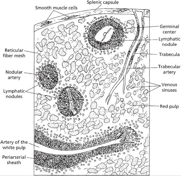
Figure 16-4. Basic internal anatomy of a spleen. (Reprinted with permission of Wiley-Blackwell from Reece, W.O. Functional Anatomy and Physiology of Domestic Animals, 3rd edition. Baltimore: Lippincott, Williams & Wilkins, 2005.)
Although the spleen is a useful organ, it is not essential in the adult, as all of its functions can be carried on by other organs. The spleen can be removed (splenectomy) without significant impairment to a mature animal.
Thymus
The thymus is an organ of immature animals, undergoing involution at puberty, although never completely disappearing.
it lies cranial to the heart, with portions extending along the trachea craniad into the ventral neck. The connective tissue components of the thymus form a loose areolar network that divides the organ into grossly visible lobules. Histology reveals a distinct cortex and medulla, both of which consist of accumulations of lymphocytes (in this location called thymocytes); it is within the thymus that embryonic lymphocytes undergo differentiation and leave to populate the many other lymphatic tissues of the body.Tonsils
In the most traditional sense, a tonsil is an unencapsulated aggregate of lymphatic nodules associated with the pharyngeal mucosa. These aggregates lack afferent lymphatic vessels, instead relying on their proximity to the epithelial surface to make contact with antigens. Many tonsils are characterized by deep invaginations on their epithelial surfaces called crypts, which presumably increase the surface area for contact with lymphatic tissue. Although the word tonsil is usually reserved for the lymphatic organs associated with the pharynx, identical histological elements are found in the mucous membranes of the prepuce and vagina and in the submucosa of the intestinal tract.