LYMPHATIC VESSELS
During lactation, the udder experiences a significant increase in blood flow, resulting in the production of a substantial volume of lymph, approximately 1.6 times the volume of milk produced.
This lymph travels through a complex network of vessels to the supramammary lymph nodes, located in the mammary fat pad above the caudal glands (Figure 22.2). While some lymph may pass through the prefemoral lymph nodes, the primary drainage route is through the supramammary lymph nodes. Efferent vessels exit these nodes, travel through the inguinal ring into the abdomen, and may pass through the deep inguinal, external iliac, or pre-temporal lymph nodes before entering systemic circulation. The lymphatic vessels draining the udder are visible superficially just under the skin, especially in high- producing cattle.In cases of udder edema, which can occur in prepartum cattle or those with mastitis, the occlusion of lymph vessels due to increased pressure or loosening of tight junctions between epithelial cells can lead to the accumulation of lymph and milk components in the interstitial space, reducing lymph clearance. In severe cases, the deep inguinal node may be palpable per rectum in cows.
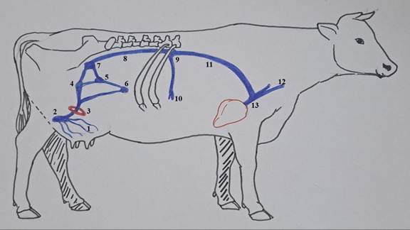
FIGURE 22.2 Diagram showing lymphatic vasculature of the mammary gland. Lymphatic vessels of the mammary gland (1); supramammary lymph gland (2); inguinal ring (3); deep inguinal lymph gland (4); external iliac lymph gland (5); prefemoral lymph gland (6); internal iliac lymph gland (7); lumbar lymph trunk (8); cisterna chyli (9); intestinal lacteals (10); thoracic lymph duct (11); jugular vein (12); anterior vena cava (13).
22.7