oints of the Appendicular Skeleton
Joints of the Thoracic Limb
The scapula has no true bony connection with the thorax. it is held in place by a number of muscles and ligaments. This type of joint is sometimes called a synsarcosis.
The shoulder (scapulohumeral) joint is spheroid. Movements in all directions, including rotation, are possible. in domestic animals, however, the arrangement of shoulder muscles practically limits movement to a hinge type of action in the sagittal plane. Thus, extension and flexion are the chief movements. The head of the humerus is a large sphere much more extensive than the comparable cavity of the scapula. The joint capsule is extensive, with poorly developed collateral ligaments. Instead, the tendons of the muscles crossing the shoulder joint on all sides act effectively as supportive ligaments (it is these well-developed tendons that blend in human beings to form the so- called rotator cuff. This term is not used in veterinary anatomy, however.).
The elbow is a true ginglymus joint formed by the humeral condyle meeting the proximal ends of the radius and ulna. The proximal end of the radius is slightly concave and expanded to give an extensive surface for support. Combined with semilunar notch of the ulna, the radius forms a half-circle embracing the humeral condyle. In the horse and ox, movement in the elbow is limited to flexion and extension. In humans and to a lesser degree in carnivores, the joint between the radius and ulna permits supination and pronation.
The carpus (Figs. 6-4 and 6-5) is a complex joint that permits flexion and extension not only between the radius and proximal row of carpal bones (radiocarpal joint), but also to a lesser degree between the proximal and distal rows of carpal bones (midcarpal joint). The entire joint is capable of absorbing considerable shock because of the many small joints formed by adjacent carpal bones connected by short ligaments.
The joint between the distal row of carpal bones and the metacarpus (carpometacarpal joint) is almost entirely a plane joint, which allows only limited gliding movements and makes almost no contribution to the degree to which the entire carpus can flex.The fibrous layer of the joint capsule of the carpus is extensive, being a long sleeve extending from the radius to the metacarpus and enclosing the carpal bones. The synovial membrane, however, forms three separate sacs: a radiocarpal sac, a midcarpal sac, and a carpometacarpal sac.
In the horse, there is normally little movement between the large metacarpal III and the smaller metacarpals II and IV (splint bones). Excessive movement or trauma results in inflammation at this site, creating a splint. In an acute state, the splint is a painful swelling where the shafts of the large and small metacarpal bones meet. Later this swelling may ossify and form a bony prominence that may not cause any lameness at all (see Fig. 4-9).
In the ox and sheep, the third and fourth metacarpal bones are fused to form the single cannon bone, which articulates proximally with the distal carpal bones and distally with the proximal phalanges. In the dog and pig, the proximal ends of adjacent metacarpal bones abut one another in a series of plane joints (intermetacarpal joints).
The metacarpophalangeal (fetlock) joint of the horse (Fig. 6-6) is formed by the distal end of the metacarpus; the proximal end of the first phalanx, or long pastern bone; and the two proximal sesamoid bones. It is a ginglymus joint that in the normal standing position is hyperextended (Fig. 6-3).
The proximal interphalangeal (pastern) joint is a ginglymus joint between the first and second phalanges (the long and the short pastern bones). Although it is a ginglymus joint, it is rather limited in motion. Degenerative changes in this joint are called a high ringbone.
The distal interphalangeal joint (coffin joint) is formed by the second and third phalanges and the distal sesamoid (navicular) bone.
The coffin joint is largely encased within the hoof and is essentially a ginglymus joint. Degenerative joint disease in this joint is commonly referred to as low ringbone. A similar pattern of articulations is followed for each digit in animals possessing more than one digit per foot (e.g., ruminants and pigs).Joints of the Pelvic Limb
The sacroiliac joint is the only bony connection between the axial and appendicular skeletons. In the young animal, this joint exhibits features of both synchondroses and synovial joints, although its mobility is progressively diminished in the adult. The articular surface of the sacrum is held in tight apposition to the wing of the ilium by a number of short, strong ligaments. Movement in this joint is normally
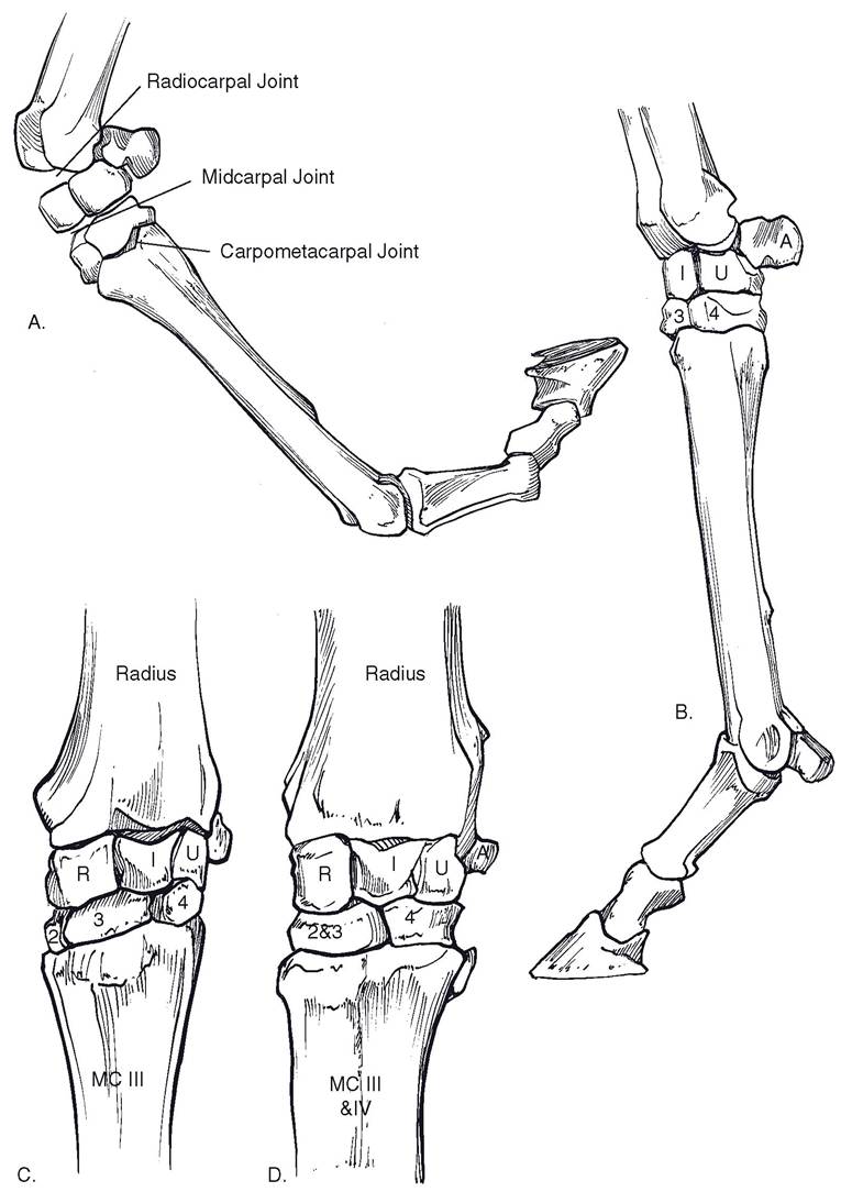
Figure 6-4. Carpus and distal joints of limb. A) Equine thoracic limb in flexion. Most of the carpal flexion derives from flexion at radiocarpal and midcarpal joints; the carpometacarpal joint has little movement. B) Lateral view of equine thoracic limb in weight-bearing position. C) Dorsal view of the equine carpus. D) Dorsal view of the bovine carpus.
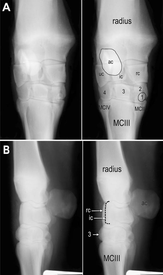
Figure 6-5. Radiographs of equine carpus. A) Dorsal-palmar projection. B) Lateral projection. MCIII = metacarpus 3; MCII = metacarpus 2; MCIV = metacarpus; rc = radial carpal bone; ic = intermediate carpal bone; uc = ulnar carpal bone; ac = accessory carpal bone. Distal row of carpal bones are numbered. Superimposed carpal bones in lateral view are not labeled. Radiographs courtesy of Susan Kraft, DVM.
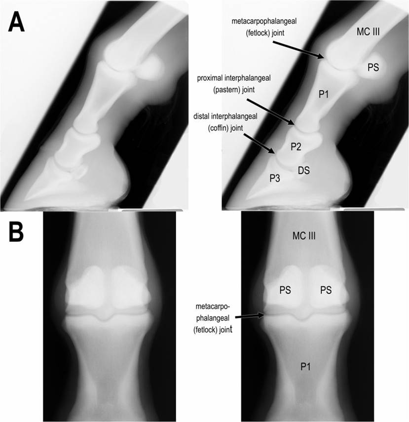
Figure 6-6. Radiographs of equine digit (a lateral projection) (A) and fetlock (a cranial-caudal projection) (B). MCIII = third metacarpal bone; P1 = proximal phalanx; P2 = middle phalanx; P3 = distal phalanx; PS = proximal sesamoid bones; DS = distal sesamoid (navicular) bone.
Radiographs courtesy of Susan Kraft, DVM.severely limited, but it may become more extensive just prior to parturition, when the ligaments stretch under the influence of the hormone relaxin (see Chapter 28). Ligaments in this area include dorsal and ventral sacroiliac and sacrotuberous ligaments. In the ox, sheep, and horse, the latter is a strong, wide band that extends from the sacrum to the tuber ischiadicum and helps form the lateral wall of the pelvis.
The sacroiliac joint can be partially separated (a sacroiliac subluxation) by a fall or other trauma. Such an injury produces pain and muscle spasm and often becomes a
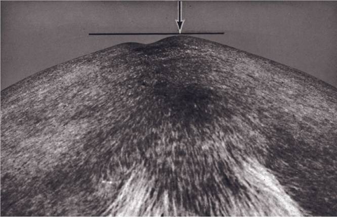
Figure 6-7. Hunter's bump, an asymmetry of the tubera sacrales produced by subluxation of the sacroiliac joint. The higher tuber sacrale is the affected side. (Reprinted with permission of Wiley-Blackwell from Stashak, T. S. Adams’ Lameness in Horses. 5th ed. Baltimore: Lippincott Williams & Wilkins, 2002.)
source of chronic soreness. Sometimes visible asymmetry in the two tubera sacrales develops, with the subluxated side displaced upward. This sign is commonly called hunter’s bump (Fig. 6-7).
The coxofemoral (hip) joint is the best example of a spheroid (ball and socket) joint. The head of the femur is about two-thirds of a sphere that fits into the less extensive acetabulum of the os coxae. The margin of the acetabulum is reinforced and deepened by a marginal cartilage.
The joint capsule of the hip joint is extensive, but not so extensive as that of the shoulder. The ligament of the femoral head (formerly round ligament) connects the head of the femur with a nonarticular area within the acetabulum. The hip joint of the horse is reinforced by an accessory ligament that extends from the prepubic tendon to the head of the femur. it is presumed to prevent significant abduction of the pelvic limb. Movements in nearly all directions are possible in the hip joint, but as in the shoulder joint, extension and flexion are the movements chiefly employed in normal locomotion.
The stifle joint corresponds to the human knee (Figs. 6-8 and 6-9). It comprises the condyles of the distal femur, the patella, and the proximal tibia. The femoral condyles are separated from the proximal tibia by two intra-articular menisci. Each meniscus is a halfmoon-shaped disk that conforms to the surface of the proximal tibia and is concave on the upper surface to fit the respective condyle of the femur. These menisci help keep the joint congruent and absorb shock. The stifle is stabilized by medial and lateral collateral ligaments and by two intra-capsular cruciate ligaments that form an X as they cross from the tibia to the femur in the middle of the joint.
The patella (kneecap) is a sesamoid bone embedded in the tendon of insertion of the large cranial muscles of the thigh. This muscle group (see Chapter 7) is a powerful extensor of the stifle, acting through its connection to the cranial aspect of the proximal tibia via one (pig) or three (horses and ruminants) strong patellar ligaments. In the horse, the medial patellar ligament is attached to the medial aspect of the
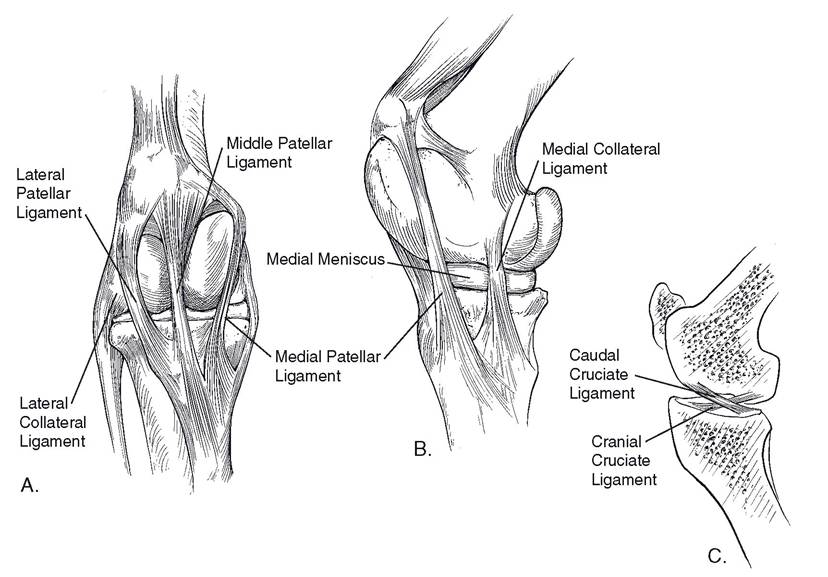
Figure 6-8. Equine stifle. A) Cranial view of right stifle. Note the medial patellar ligament's relationship to the medial ridge of the femoral trochlea and the presence of the medial and lateral menisci within the femorotibial articulation. B) Medial view of right stifle. In this position the patella is locked over the trochlea and no muscular effort is required to keep the joint extended. C) A sagittal section of the stifle showing the cruciate ligaments, whose intra-articular location provides cranial-to-caudal stability to the joint.
patellar via a large, hook-shaped fibrocartilage. The combined cartilage and tendon create a stout loop that can be locked over the medial ridge of the femoral trochlea at will (Fig. 6-8). In this position, the stifle is held in extension with minimal muscular effort; this anatomic arrangement therefore contributes to the ability of the horse to stand while sleeping.
The tarsus (hock) joint, like the carpus, is a composite joint (Figs. 6-10 and 6-11). The ginglymus portion is formed between the distal end of the tibia and the talus. This portion of the joint is held together by strong medial and lateral collateral ligaments of the hock.
The calcaneus projects proximad and caudad to form a lever for attachment of the common calcaneal tendon (Achilles tendon), which is the common insertional tendon of the extensor muscles of the hock. The calcaneus is firmly attached to the other tarsal bones by many short, strong ligaments. The ligaments are less extensive over the craniomedial aspect of the hock. In this location, the joint capsule is immediately beneath the skin, and distension of this joint results in an obvious soft bulge commonly called a bog spavin.. In the horse, movement between adjacent tarsal bones is limited to a very small degree of gliding. However, in the ox, sheep, and pig, the proximal intertarsal joint has some hinge movement. Distal to the hock, the joints are similar to those of the forelimb.
Figure 6-9. Lateral radiograph of equine stifle joint. Radiographs courtesy of Susan Kraft, DVM.
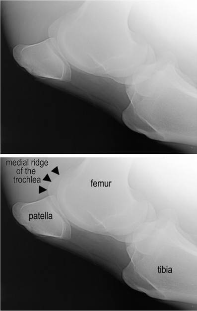
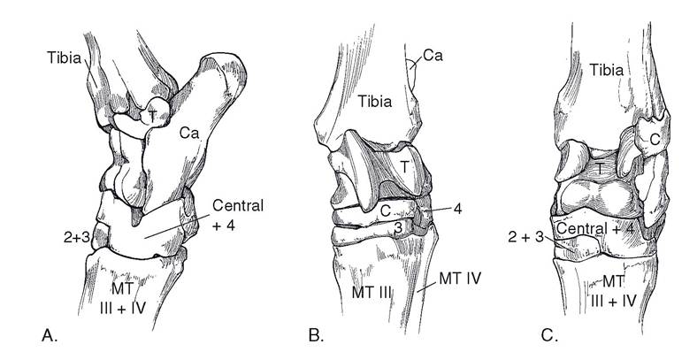
Figure 6-10. Hock. A) Lateral view of left bovine hock. B) Cranial view of left equine hock. C) Cranial view of left bovine hock.
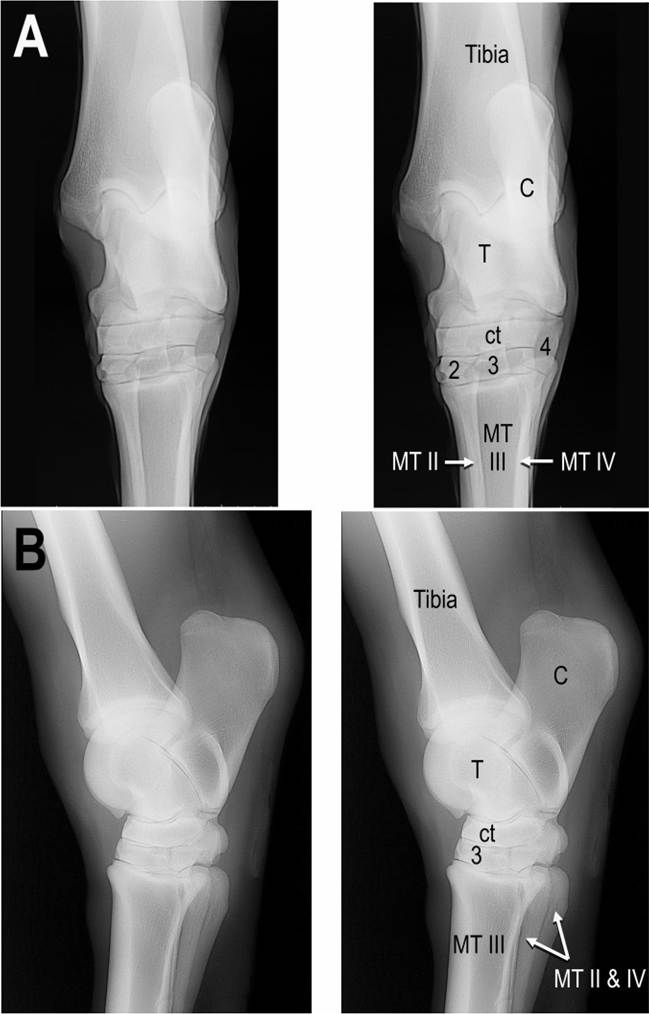
Figure 6-11. A) Cranial-caudal projection of the equine tarsus. B) Lateral projection of the equine tarsus. C, calcaneus; T, talus; ct, central tarsal bone; 2, second tarsal bone; 3, third tarsal bone; 4, fourth tarsal bone; MT III, third metatarsal bone; MT II, second metatarsal bone (medial splint bone); MT IV, fourth metatarsal bone (lateral splint bone). The second and fourth tarsal bones are largely superimposed in the lateral view and are therefore not labeled. Radiographs courtesy of Susan Kraft, DVM.