Olfaction
Key points
■ Odoriferous substances stimulate olfactory neurons. Action potentials pass caudally into the cranial vault, synapsing in the olfactory bulb. Olfactory tract axons project to both sides of the forebrain for olfactory perception, stimulation of behavioural responses and to the brainstem for reflex function.
■ Input from the vomeronasal organ is via the olfactory nerve and may stimulate the Flehmen reaction during sexual activity in some species.
The name olfactory ‘nerve’ is actually a misnomer since it consists entirely of CNS tissue (see p1), however in humans the olfactory bulb is so diminutive as to resemble a nerve. In veterinary species, the olfactory bulb is prominent (Figs. 10.3, A2, A3, A10).
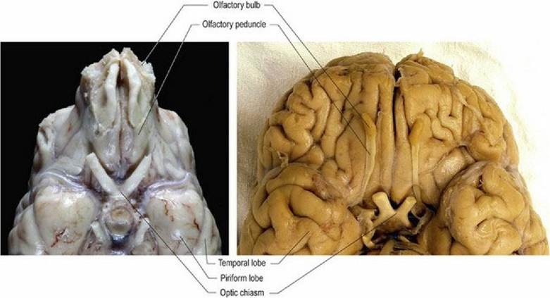
Fig. 10.3 Comparison of the olfactory bulbs of the dog (macrosmatic) and human (micromatic) brains,
ventral aspects.
(Image of human brain courtesy of Dr. Henry Waldvogel, University of Auckland.)
The olfactory mucosa is located on the ethmoidal labyrinth and dorsal nasal septum in the dog. Grossly, the olfactory mucosa may be slightly yellowish compared with surrounding mucosa, due to pigment in the sustentacular cells. Afferent fibres from the olfactory mucosa and vomeronasal organ contribute to CN I, which is unmyelinated. Olfactory neurons are bipolar cells with 6-8 long cilia that project into the overlying mucus in the caudodorsal part of the nasal cavity. Olfactory receptors are also located in the vomeronasal organ on the rostral floor of the nasal cavity. These receptors may respond to pheromones, which are chemicals that trigger social responses between members of the same species. The vomeronasal organ may mediate the curling of the upper lip, the Flehmen reaction, in males scenting females with respect to mating suitability.
Odoriferous substances dissolve in the mucus overlying the olfactory mucosa, or in the vomeronasal organ, stimulating the olfactory neurons. As odoriferous substances can be cytotoxic, olfactory neurons can be renewed from stem cells located at the base of the olfactory mucosa. The potential for harvesting olfactory stem cells and using them as a source of new neurons for a patient is currently an area of active research in neuroscience.Axons from the olfactory mucosa and the vomeronasal organ pass through the cribiform plate into the cranial vault and synapse on the olfactory bulb neurons. Post-synaptic axons travel caudally in the olfactory
tract of the olfactory peduncle. The tract splits into lateral, intermediate and medial stria (see Fig. A11). The axons of the lateral stria synapse in the olfactory tubercle and pass to the cortex of the piriform lobe for olfactory perception. Fibres also connect to the limbic system (see Chapter 11). More medial fibres pass to the septal nuclei, which are located between the rostral aspects of the lateral ventricles, and to the hypothalamus and reticular formation of the brainstem. Fibres also decussate via the rostral commissure and pass to the contralateral olfactory bulb. The rostral commissure also interconnects the two piriform lobes. Through the connections to the limbic system, cerebral cortex and hypothalamus, olfaction can be a potent stimulator of behaviour and emotional states. Connections to the brainstem permit olfacto-visceral reflexes such as salivation in response to olfactory stimulation (Fig. 10.4).
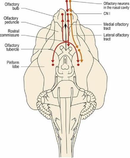
Fig. 10.4 Canine brain, ventral aspect, olfactory pathways and their connections.
Note that olfaction does not pass through the thalamus; this is different to all other afferent information which does pass through the thalamus en route to the cerebral cortex.
Lesions in one olfactory bulb lead to unilateral anosmia; this is hard to detect clinically. Bilateral lesions are required in the olfactory mucosa, or bulbs, peduncles or piriform lobes to cause complete anosmia.