REPRODUCTION IN THE AVIAN FEMALE
1. What is the oviduct in the avian female?
2. Is the avian reproductive tract bilateral? Which side persists?
3. What are the component parts of the oviduct?
4. What are the functions of the infundibulum?
5.
What is the function of the magnum?6. What is the function of the isthmus?
7. What are the functions of the uterus? What is the cuticle, and what is its function?
8. What is the function of the vagina?
9. What are sperm-host glands?
0. Where are the yolk proteins and lipids formed?
11. What is the function of the chalazia?
2. What is the immediate source of the calcium needed for hard shell formation?
3. What is the length of an ovulation cycle in the domestic hen? What is a clutch?
4. What is meant by “egg bound” in the avian female?
L5. Describe oviposition in the avian female.
The term oviduct is the anatomic term used to describe the complete tubular genitalia of the avian female. It is highly coiled and extends from the ovary to the cloaca. In the sexually mature chicken, it can be straightened out to a length of 70 to 80 cm. With few exceptions, among the domestic species of birds, only the left ovary and oviduct reach functional development. The left ovary is cranial to the left kidney and is tightly attached to the dorsal body wall, caudal to the left lung, and adhered closely to the caudal vena cava.
The oviduct can be subdivided into five functional regions (Figure 15-27). Beginning with the ovarian end and extending to the cloaca, they are the infundibulum, magnum, isthmus, uterus (shell gland), and vagina. As described in Chapters 10 and 11, the cloaca is a region through which digestive and kidney wastes and genital tract products pass. The function of the infundibulum is to envelop the ovulated oocyte with its yolk and begin its direction through the remaining portions of the oviduct.
The infundibulum is also the location where fertilization would occur because it is assumed that spermatozoa would not be able to penetrate the oocyte after it begins to be covered by albumen (egg white). Secretion of albumen occurs in the magnum and it is the longest segment of the oviduct. Albumen surrounds the central yolk mass and constitutes about two-thirds of the egg’s weight. The isthmus secretes the fibrous inner and outer shell membranes that enclose the contents of the egg and provide support for deposition of the hard shell. The uterus adds fluid to the developing egg, secretes the hard shell, and adds the cuticle (a proteinaceous layer) to the exterior of the hard shell. The cuticle functions as a blockade against the entrance of bacteria and reduces water loss. The uterus is also the location where pigment is added to the hard shell (e.g., brown eggs). The uterovaginal sphincter, a constricting muscle, is the dividing point between the uterus and vagina. The oviduct terminates with the vagina that is attached to the cloaca. Nothing is added to the egg as it moves through the vagina; its function seems to be the prolonged storage of spermatozoa in its sperm-host glands (additional sperm-host glands of the chicken, but not turkey, are located in the infundibulum). Storage and maintenance of fertilizing capacity for spermatozoa is possible in these glands for 7 to 14 days for chickens and 40 to 50 days for turkeys. A summary of the hen’s egg formation is provided in Table 15-4.| TABLE 15-4 HEN’S REPRODUCTIVE TRACT AND EGG FORMATION | |||
| OVIDUCT SEGMENT | LENGTH IN cmα | GENERAL FUNCTION | TIME SPENT |
| Infundibulum | 7-8 | Capture ovulated ova Fertilization | 15 min |
| Magnum | 30 | Albumen production | 3 h |
| Isthmus | 10 | Shell membranes created | About 1 h |
| Uterus (shell gland) | 12 | Addition of fluid (plumping) and albumen layering Egg shell production | 20 h |
| Vagina | 12 | Egg transport Sperm storage | 1 min |
| “Segment lengths vary greatly depending on relaxation and contraction. The lengths reported are for an active reproductive tract producing eggs. Data from: Burke WH. Avian reproduction. In: Swenson MJ, Reece WO, eds. Dukes’ Physiology of Domestic Animals. 11th edn. Ithaca, NY: Cornell University Press, 1993. | |||
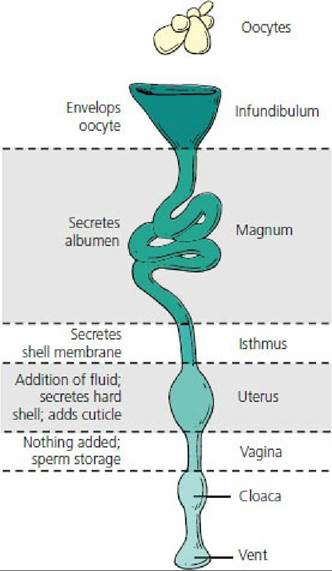
■ FIGURE 15-27 The five functional regions of the oviduct of the laying hen. The oviduct is the complete tubular genitalia of the avian female and consists of the infundibulum, magnum, isthmus, uterus, and vagina.
As in mammals at the time of birth, only a small number of the ovarian follicles present at the time of hatching develop to the point of ovulation. The immature avian follicle consists of an oocyte surrounded by granulosa cells and proceeds to the mature follicle, which is quite large because of the addition of yolk material. The majority of yolk materials are deposited into the follicle during its rapid growth phase (final 7 to 11 days before ovulation). Yolk protein and lipid formation occur in the liver and are transported via the blood to the ovary. Deposition of yolk into the maturing follicle terminates about 24 hours before ovulation. Yellow egg yolk is a complex mixture of water, lipid, protein, and many components in very small amounts, including vitamins and minerals. Inasmuch as there will be no maternal source of nutrition, the yolk is the nutritional source for the developing embryo. The yellow color of egg yolk is caused by xanthophyll pigments in the diet. It is possible to have light yellow or white egg yolks when xanthophyll is low or lacking in the diet.
A cross-section of a hen’s egg is shown in Figure 15-28. There are four distinct layers of albumen in the laid egg: (1) the chalaziferous layer (thick), attached to the yolk; (2) the inner (liquid) layer; (3) the dense (thick) layer; and (4) the outer (thin) layer. Extending away from the yolk toward both ends of the egg are twisted strands of protein called the chalazae.
They are extensions of the chalaziferous layer. It has been suggested that the function of the chalazae is to hold the yolk and developing embryo in the center of the egg so that adhesion of the embryo to the shell membranes does not occur. Most of the shell is secreted during the last 15 hours that the egg spends in the uterus. Its major composition is calcium carbonate. All of the calcium secreted into the uterus during shell formation comes from the blood. Because the amount of calcium in the shell exceeds the total amount of calcium in the blood, a dynamic interchange of skeletal calcium with blood calcium must occur. A sizeable amount of each shell’s calcium is therefore derived from the bones. Blood and bone calcium is replaced by dietary calcium.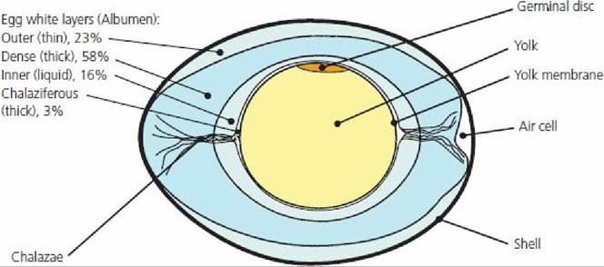
■ FIGURE 15-28 Midsagittal section of a hen’s egg. The chalazae are extensions of the chalaziferous layer that hold the yolk and developing embryo in the center so that adhesions of the embryo to the shell membranes do not occur. The approximate percentage of the albumen for each layer is shown.
The ovulatory cycle in the domestic hen is about 24 to 26 hours long and cycles may be repeated day after day without interruption. The period of time from one interruption to the next is called a clutch. In chickens, the clutches can range from one to 30 or more eggs. Oocytes not enveloped by the infundibulum are reabsorbed. Occasionally, an atypical large egg may become lodged in the lower end of the shell gland or in the cloaca. The condition is referred to as “egg bound” and if not relieved can lead to death of the bird.
The act of laying of the egg is known as oviposition. The muscles of the shell gland contract and the sphincter separating the shell gland from the vagina relaxes. The shell gland contractions coordinate with abdominal muscle contractions (bearing-down reflex) to expel the egg.
Some commercial flocks of egg-laying hens begin to lay at about 22 weeks of age and continue for about 1 year. In this case an average production of about 260 eggs per hen per year is typical. Those breeds of chickens that are selected for meat production lay far fewer eggs.
Aside from those breeds selected for egg production, there is a tendency among turkeys and breeds of chickens selected for meat production to show broodiness, an incubation behavior in birds. After a few weeks of laying eggs, there is a desire to incubate them by setting on them in a nest. When hens become broody, the ovaries regress and egg production ceases. The hormone prolactin causes the ovarian and behavioral changes associated with broodiness.
■