REPRODUCTIVE SYSTEM
bloodstream to the pituitary gland. In birds two forms of GnRH are released but the exact demarcation of functions is still under research (Millam 1997). This then stimulates the production of follicle-stimulating hormone (FSH) and lutropin luteinizing hormone (LH), which trigger gonado- genesis and breeding (Kirby & Froman 2000).
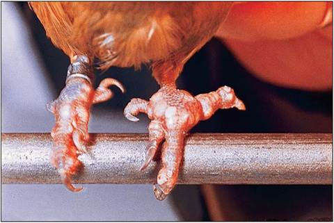
Figure 6.58 • Articular gout in a budgie showing dorsal nodules of white crystalline tophi.
Reproduction in the bird follows a definite breeding cycle that is controlled by environmental factors like photoperiod, food availability, and temperature. In temperate climates the trigger factor is the increasing daylight of spring whereas in arid climates the advent of the rainy season bringing increasing food supplies stimulates the gonads to develop (Millam 1997). In order to minimize weight the gonads enlarge for the breeding season and become small and dormant at the end of the season (Evans 1996). In some species this can be dramatic; for example, the gonads of the starling (Sturnus vulgaris) increase 1500 times in size during the breeding season (Maina 1996).
The pineal gland is thought to be the center of the complex reproductive and migratory avian clock. However, the main detector of increasing daylight lies not in the eyes or pineal gland but in the hypothalamus. Here photoreceptors release gonadotropin-releasing hormone (GnRH) via the
Once breeding is finished the shorter days of summer stimulate resorption of gonadal tissue and allows time for molting. Under the influences of prolactin and the pineal gland migratory species lay down fat and increase food intake. The short days of winter then inhibit the gonads, allowing them to be stimulated again in the spring.
Birds differ from mammals in that the female is het- erogametic, being ZW, and the male is homogametic, being ZZ.
This means the sex of future offspring is decided after ovulation and not after fertilization.SEXING BIRDS
Many birds and especially psittacines are monomorphic, making it difficult to distinguish gender. The following methods can be used:
• External secondary sexual characteristics - Fowl have some useful sex-linked color/feather traits exploited by commercial chicken farms for rapid sexing. Male budgies have a blue cere (Fig. 6.59) while the hen has a brown cere. The male Eclectus parrot is vivid green but the hen is mainly red.
• Vent sexing - This was the traditional method used in poultry farming. Day old chicks were sexed by everting the lip of the vent to expose this tiny genital region. This appears rounded in the male chick and conical in females.
• Surgical sexing - This involves direct visualization of the gonads by laparoscopy into the left abdominal airsac under anesthetic.
• DNA analysis of blood.
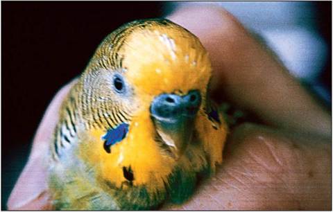
Figure 6.59 • Cock budgie (Melopsittacus undulatus) showing blue cere.
Male
Testes
The bean-shaped testes are paired and lie near the cranial pole of the kidney, just caudal to the adrenal glands. Medially, the testes lie close to the aorta and the caudal vena cava. Each testicle is suspended by a short mesorchium and is surrounded medially by the abdominal airsac. The left testis tends to be larger than the right in immature birds (King & McLelland 1984) (Fig. 6.60).
The testes are under the influence of FSH and LH and the dimensions can increase dramatically with sexual activity. In the non-breeding season the testes shrink to almost nothing and may be hard to visualize. The dormant testes
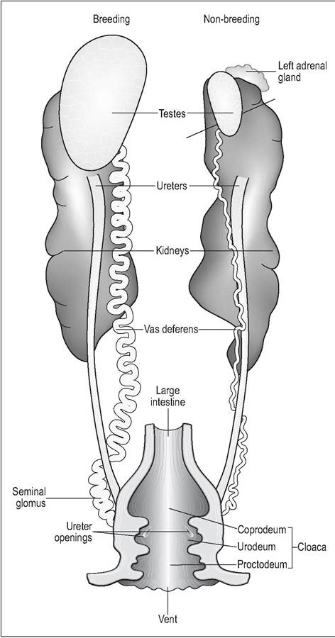
Figure 6.60 • Ventral view of male passerine urogenital tract. The right side shows the large breeding testes and seminal glomus while the left side shows the dormant non-breeding testes.
are light brown to yellow in color, turning white when the bird becomes sexually active. In some psittacine species, like cockatoos (Cacatua spp.) and macaws (Ara spp.), the immature or dormant testes can even appear black due to melanocytes located in the interstitium of the testes.
The testes consist of seminiferous tubules, which produce the sperm from the epithelium. The interstitial or Leydig cells lie meshed between the seminiferous tubules and, under the influence of LH, produce the main androgens: testosterone and androstenedione (Kirby & Froman 2000; Lake 1981). These hormones provide the secondary sex characteristics like coloration and song during courtship.
The tunica albuginea is much thinner than in mammals and there is no pampiniform plexus. The epididymis is smaller and less developed as sperm maturation occurs in the vas deferens and not, as in mammals, in the epididymis. The vas deferens is closely associated with the ureter in the dor- somedial midline celom but is distinguished from it by its zig-zag appearance. It enters the dorsal wall of the urodeum. There are no accessory sexual glands.
CLINICAL NOTE
In domestic fowl the epididymis has an appendix which attaches by connective tissue into the ventral part of the adrenal gland. Surgical castration is therefore not always permanent because in castrated males this area of tissue can sprout nodules that secrete androgens (King & McLelland 1984).
HOW CAN SPERM DEVELOP AT THE HIGH AVIAN TEMPERATURE OF 40-42° C?
• Most spermatogenesis occurs in the early hours of the morning when temperatures are coolest.
• The adjacent abdominal airsacs may play a role in cooling (Maina 1996).
• In passerines, which have the highest body temperatures, the vas deferens elongates distally to form a cloacal promontory called the seminal glomus. This is the main site of sperm storage and functions like a scrotum in keeping the sperm at temperatures 4° C lower than
the core temperature.
The seminal glomus projects into the cloaca and helps sexually differentiate these species during the breeding season (Lake 1981; Orosz et al. 1997).may contribute to the seminal fluid (Lake 1981). Sperm remains viable in the female tract for much longer than in mammals and may survive for 5 or 6 days.
Absence of phallus
Psittacines, passerines, pigeons and birds of prey all have no phallus. These species copulate by transferring semen from the everted cloaca directly into the female oviduct (King, AS 1981b).
Non-protrusible phallus
A rudimentary non-protrusible phallus is seen in male turkeys and chickens and lies on the ventral lip of the vent. It consists of a small median tubercle intimately associated on each side with lymphatic folds and vessels. When erected with lymph the phallus develops a median groove which permits passage of ejaculate down into the everted female oviduct (King, AS 1981b; King & McLelland 1984).
Protrusible phallus
The protrusible phallus is elongated and capable of true intromission into the female cloaca and is seen in ratites and Anseriformes. The latter have a curved fibrous phallus that conveys semen via a spiral groove. The distal end lies enclosed in a cavity on the floor of the cloaca and becomes engorged with lymphatic fluid (Fowler 1986; King, AS 1981b).
KEY POINTS
• Size and color changes between dormant and active testes
• Epididymis less well developed. Its close connection to adrenal gland in chickens makes permanent castration difficult
•Seminal glomus used for sperm storage in passerines
•Most birds lack a true phallus
Female
The female embryo has two gonads but only the left one develops, leaving the right ovary and oviduct to regress. This can be identified as a strand of tissue on the right side along the ventral side of the caudal vena cava (Gilbert 1979). As in the male, the female organs regress dramatically after the breeding season (King & McLelland 1984).
Phallus
When present, the avian phallus is solely reproductive and becomes engorged by lymph fluid instead of blood during erection (Kirby & Froman 2000).
Owing to the lack of accessory sex glands, avian semen has low volume (e.g., the cockerel has an ejaculate of only 0.5-1 ml) but some lymphCLINICAL NOTE
The right ovary is sometimes retained in birds of prey, although it is rare for it to have a functional right oviduct. Other species like the Common kiwi (Apteryx australis) have been known to have two ovaries (Gilbert 1979; King & McLelland 1984).
Sexual maturity
Most domestic fowl come into lay at around 5 months. Seasonal birds will lay in the first spring after hatching. Japanese quail (Coturnix coturnix) become sexually active at 5-6 weeks, which is why they are often used in laboratory research.
In ducks, geese, and swans the oviduct is covered by a small membrane where it opens into the urodeum, until the bird reaches sexual maturity. This can be used to distinguish juvenile from mature birds.
Ovary
The left ovary lies caudal to the adrenal gland and near to the cranial tip of the kidney. It consists of a vascular medulla, with nerve fibers and smooth muscle, and a peripheral cortex. It is suspended by the mesovarium and receives its blood supply from the cranial renal artery.
The ovary resembles a bunch of grapes due to large follicles in sexually active hens (Gilbert 1979). The follicle is suspended by a stalk containing smooth muscle that has a rich vascular and nerve supply.
CLINICAL NOTE
The cranial renal artery is very short, making ovariectomy very difficult and bringing a high risk of hemorrhage.
Prevention of excessive egg laying is done instead by removal only of the oviduct (salpingohysterectomy) (Orosz et al.
1997).
Oviduct
The oviduct occupies the left dorsocaudal side of the celomic cavity (Fig. 6.61). It is a long coiled tube suspended by the mesosalpinx. The wall consists of ciliated epithelial lining, glands, and smooth muscle. The smooth muscles are thickest in the uterus and vagina to aid with sperm transport. It is divided into five parts: the infundibulum, magnum, isthmus, shell gland (uterus), and vagina (Gilbert 1979; King & McLelland 1984).
Infundibulum
Unlike mammals, fertilization is not a prerequisite for egg formation and development. The funnel of the infundibulum catches the egg, and fertilization must take place here before the oocyte gets surrounded by albumin (Johnson, AL 2000). Penetration by sperm usually occurs within 15 minutes of ovulation. A thin and dense layer of albumin is then added in the tubular section to surround the yolk. Some species have a sperm host gland in this area to store sperm for a variable time, for later fertilization.
Magnum
The magnum is the longest part of the oviduct; it is coiled and numerous tubular glands give it a thickened appearance.
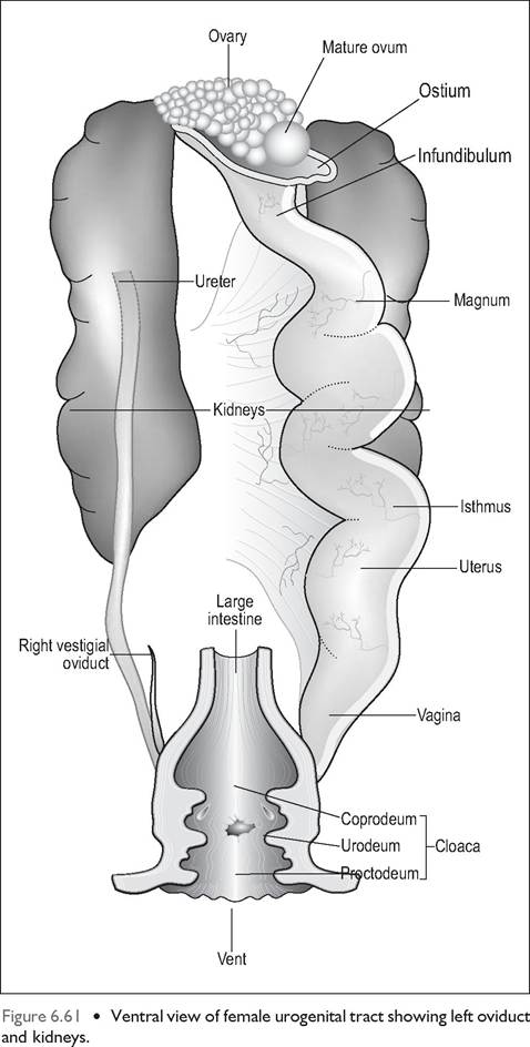
These glands produce the egg white protein albumin, which gives the lumen a milky white color. Calcium, sodium, and magnesium are also added here.
Isthmus
The isthmus divides the magnum from the uterus and is present in poultry but not in psittacines (Orosz et al. 1997). The folds are less prominent than the magnum but its gland secretes more albumin.
Uterus
This holds the egg during shell formation and the majority of time (80%) is spent here (Gilbert 1979). The uterus is very vascular to aid with calcium deposition.
Vagina
The vagina is separated from the uterus by a vaginal sphincter. The smooth muscle here is more powerful than the rest of the oviduct. In some species the egg can remain here for hardening before the egg passes out of the oviduct into the urodeum.
Sperm host glands are in the uterovaginal junction where sperm can be stored for many months, which is why female birds can be fertile weeks after the absence of the male (Johnson, AL 2000).
Endocrine control
In females FSH is mainly responsible for follicular growth. The developing follicles produce estrogen from the theca and interstitial cells, and progesterone from the granulosa cells. Increasing levels of estrogen stimulate a LH surge and, under the influence of this, the follicle splits to release the primary oocyte. Estrogen also mobilizes calcium from the bone, increasing plasma calcium for egg formation.
Progesterone continues to be secreted to inhibit further ovulation and stimulate behavioral changes associated with broodiness and incubation. Prolactin stimulates the production of crop milk in both male and female pigeons. Oviposition in birds is controlled by prostaglandins and arginine vaso- tocin/oxytocin.
Ovulation
The number of ovulations varies with the species but most wild birds lay only one clutch a year. The infundibulum catches the oocyte and this is facilitated by the left abdominal air sac, which tightly encloses the ovary (Gilbert 1979). However, if the sequence gets out of phase oocytes can be lost into the celom. Some of these will get absorbed but others may cause egg peritonitis. Once ovulation has taken place the follicle shrinks and regresses. No corpus luteum remains as there is no developing embryo to maintain.
GENERAL INTEREST
The avian primary oocyte is the largest animal cell.
The oocyte of the now extinct Madagascar Elephant bird (Aepyornis maximus), thought to be the origin of the mythical Roc, had a diameter of 37 cm, and the volume of a bucket!
Egg formation
In the chicken the developing egg travels down the oviduct in about 25 hours, but it takes about 48 hours in psittacines. Protein and lipid are synthesized in the liver and travel to the oocyte in the ovary where they are made into yolk (vitellogenesis). In the infundibulum a thin layer of albumin is laid down during the short 15 minute passage. It takes 3 hours to go through the magnum where it acquires albumin, sodium, calcium, and magnesium.
In the isthmus the inner and outer shell membranes are formed and calcification begins. The last 20 hours are spent in the uterus where the shell is formed and the albumin doubles in volume, creating the “plumping effect”. During the last 15 hours the highly vascular uterus draws calcium rapidly from the blood. The completed egg then passes through the vagina in seconds to be expelled from the vent (King & McLelland 1984).
Oviposition
Expulsion of the egg involves contraction of the uterus (shell gland) with relaxation of abdominal muscles and can take from a few hours to minutes. Cuckoos have the ability to lay their small eggs in a few seconds so they can get away before the host returns (Gilbert 1979; King & McLelland 1984). Contraction of the uterus is thought to be controlled by a combination of vasotocin/oxytocin and prostaglandins (Johnson, AL 2000).
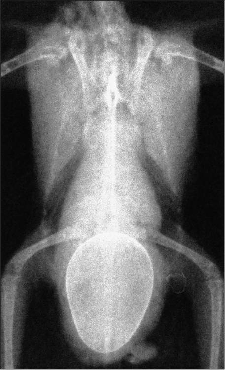
Figure 6.62 • Ventrodorsal radiograph of eggbound budgie with increased radiopacity of the pelvic limbs. In laying birds calcium is laid down in the medullary cavity prior to laying in a phenomenon called polyostotic hyperostosis. This bird also has an enlarged hepatic silhouette most likely due to hepatic lipidosis.
CLINICAL NOTE
Prostaglandins are of more use clinically for treatment of eggbound birds because they not only cause contraction of the oviduct but relax the uterovaginal sphincter.
Post egg laying
The bird enters the non-breeding state while it is incubating and caring for its brood. The resting ovary has a shrunken appearance similar to a juvenile ovary and the oviduct regresses into an inconspicuous, narrow tube.
GENERAL INTEREST
The Red jungle fowl can lay eggs indeterminately and can quickly lay more if eggs are lost from the clutch. Domestic fowl have been bred from this species and this is why they can lay eggs almost daily for up to 352 days per year.
Avian eggs
Avian eggs differ from reptilian eggs in that the principal stored nutrients are fats in the yolk sac. This produces more energy and water than protein and allows birds to survive in more arid environments. They are also porous to allow for gas exchange. The size of the egg varies with the type of newborn: altricial species lay much smaller eggs than precocial ones.
The egg consists of a germinal disc, yolk, yolk membranes, albumin, and shell (Fig. 6.63). The germinal disc is either a blastoderm (fertilized) or blastodisk (unfertilized). The yolk is thick and viscous and forms the main nutrient for the embryo. White yolk is mainly protein with some fat while yellow yolk is the reverse. The yolk membranes form a barrier between yolk and the albumin but it is permeable to water and salts.
The albumin is less viscous than the yolk and composed mainly of protein. A thin layer of albumin encloses the yolk membranes and this suspends the yolk in the center of the egg by twisted strands called chalazae.
The shell consists of the shell membranes, the testa and the cuticle. The testa is the main thickness of the shell and consists of a matrix of fibers and calcium carbonate. The cuticle is water repellent and acts as a barrier to infection. In contrast to reptile eggs, avian eggs can be pigmented by two pigments, porphyrin and biliverdin, which are deposited throughout the testa. Small flaws between the calcium carbonate crystals form pores which allow the egg to breathe (Gilbert 1979; King & McLelland 1984).
Chicks
Chicks fall into two categories: precocial (nidifugous) and altricial. The precocial chicks have natal down, hatch with their eyes open, and can survive outside the nest within 1or 2 days (Fig. 6.64). In contrast, altricial chicks are born blind and naked and require long periods of feeding.
KEY POINTS
• Birds have only a left ovary and oviduct.
• Sperm storage is possible in sperm glands in the oviduct.
• Oviposition is controlled by prostaglandins and oxytocin/ vasotocin.
• Avian eggs differ from those of reptiles in that they can be pigmented, are more porous, and the principle nutrient to the embryo is egg yolk.
• Young can be either precocial or altricial.
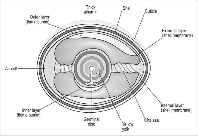
Figure 6.63 • Internal structure of the egg.
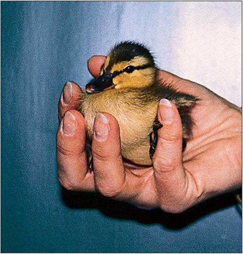
Figure 6.64 • Precocial (nidifugous) chicks are born with natal down and their eyes open and can survive outside the nest within 1-2 days.