SKELETAL SYSTEM AND INTEGUMENT
How can a reptile with a heavy shell submerge in water and walk along the bottom?
This is achieved by large lung volume, specific gravity, and stored water.
Semiaquatic turtles, like the Red-eared slider (Trachemys scripta) have a specific gravity lighter than water, so they float.
Their shell accounts for about 75% of their mass so they also have a larger pulmonary volume and extra buoyancy from their bladder and cloaca. When they dive they expel gas from their lungs to submerge (Wood & Lenfant I976).Fully aquatic turtles tend to have a specific gravity slightly greater than water and so they tend to sink and require little effort to stay underwater. They also tend to have flatter shells and less lung volume (Seymour I982).
and can remain submerged for long periods using their long necks to grab at passing prey. They have a leathery, flat shell, large webbed feet, and reduced plastron to allow for free limb movement and little water resistance (Evans 1986; Pough et al. 2002).
Snapping turtles (Chelydridae) have such a reduced shell that the head can only be retracted into the neck folds, leaving the nose visible (Fig. 3.5). These are not good swimmers and often walk along the bottom of ponds. They are omnivores and have strong jaws for predation and protection (Evans 1986; Pough et al. 2002).
Locomotion
Terrestrial chelonian species have limbs that project sideways so the muscles tire easily and progress can be laborious. Despite their awkward gait and the weight of the shell they can, however, move at great speed. Aquatic species have evolved different methods of locomotion. Freshwater turtles swim with alternate beats in a paddling motion and some can walk along the bottom. Marine turtles move their forelimbs in unison like flippers, using their rear limbs like rudders (Walker 1973).
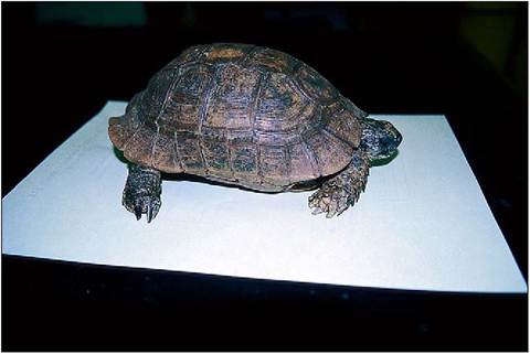
Figure 3.2 • Spur-thigh tortoise (Testudo graeca) showing the high domed shell and short, stubby legs of terrestrial species.
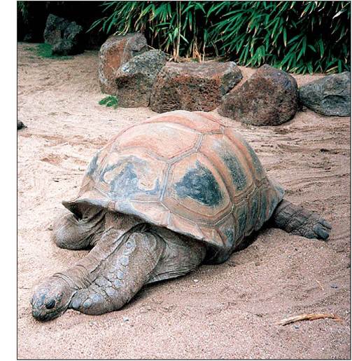
Figures 3.3 • Despite its enormous size the Galapagos tortoise (Geochelone nigra) has retained a head small enough to retract inside the shell.
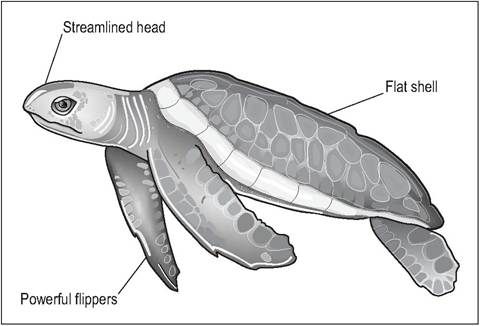
Figure 3.4 • Green sea turtle (Chelonia mydas). Marine species have evolved a flatter, softer shell for streamlining with long, fore flippers for powerful propulsion.
The shell
The dome of the shell is called the carapace and the flat underpart is called the plastron. The joint between the carapace and plastron is called the bridge. The cranial aperture is called the axillary aperture, and caudally is the inguinal aperture. The shell is formed from dermal bone and consists of about 60 bones formed from the modified pectoral and pelvic limb girdles, trunk vertebrae, sacrum, and ribs. These are covered by keratinized epidermal scales known as scutes (Fig. 3.14). These scutes do not correspond with
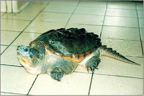
Figure 3.5 • Snapping turtle (Chelydra serpentina). The powerful snapping jaws compensate for the inability to retract the head into the shell.
the underlying bone, adding greater protection and strength to the shell (Pough et al. 1998a; Zangerl 1970).
Skeleton
Skull
Chelonians belong to the subclass Anapsida (without arches) because they lack true temporal openings (Fig. 3.6). However, many species do have gaps in the temporal region that provide a pseudotemporal fossa for muscle attachments.
Although the head has to be kept small to enable it to be retracted, the sturdy skull, large adductor muscles, and short jaw still enables chelonians to have a strong bite.
On either side of the small brain case there are large paired supratemporal fossae. Strong retractor muscles extend from these fossae and supraoccipital crest to the base of the neck, enabling them to retract their head.
These muscles also enable them to pull at food with their heads while holding it with their limbs (Evans 1986).In order to keep the head small yet retain a strong bite, the adductor muscles run through a trochlear pulley, which lengthens the muscle fibers and gives them extra strength (Fig. 3.7). In Pleurodira this pulley is formed by a process on the pterygoid bones while in Cryptodira it is formed by the quadrate bones. In each case, although the muscles originate from the back of the skull the muscle is redirected vertically for maximum force (King 1996; Pough et al. 1998a).
Mandible
In Chelonia the mouth opens by lowering the jaw (the reverse of crocodiles). Like lizards, they have a mandibular symphysis, and jaw articulation is between the quadrate bone and the articular bone of the lower jaw.
Vertebrae
There are 18 presacral vertebrae, consisting of 8 cervical and 10 trunk vertebrae. The trunk vertebrae each have rib
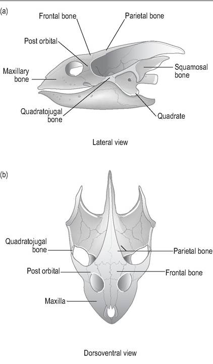
Figure 3.6 • Modified anapsid skull of chelonian.
(a) Lateral view
(b) Dorsoventral view
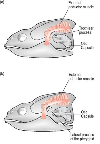
Figure 3.7 • Lateral view of chelonian skull showing adductor muscle pulley system. Redirecting the muscles vertically for maximum force enables the head to be kept small so that it can be drawn in for protection from predators.
(a) Cryptodires - the pulley runs by the quadrate bone
(b) Pleurodires - the pulley runs along the lateral process of the pterygoid bone
attachments which fuse with the dermal bone plates (Figs. 3.8-3.11). There is no sternum. In contrast to the fused trunk, the cervical and caudal vertebrae are free. The neck and tail are highly flexible and have well-developed epaxial and hypaxial muscles (Evans 1986).
Cervical vertebrae
The eight cervical vertebrae allow for bending of the neck sideways (Pleurodira) or inside the shell (Cryptodira).
The Pleurodira cannot retract their head entirely inside the shell and this lack of protection may explain why they have been less successful and their range limited to the southern continents.CLINICAL NOTE
Chelonia are the only tetrapods that have their pectoral and pelvic girdle inside their “ribs”. This means that chelonians do not possess an expandable rib cage for breathing.
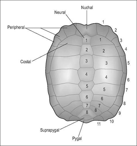
Figure 3.8 • Dorsal carapace showing dermal bone plates.
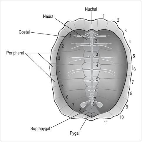
Figure 3.9 • Ventral carapace showing dermal bone plates.
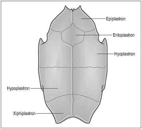
Figure 3.10 • Ventral plastron showing dermal bone plates.
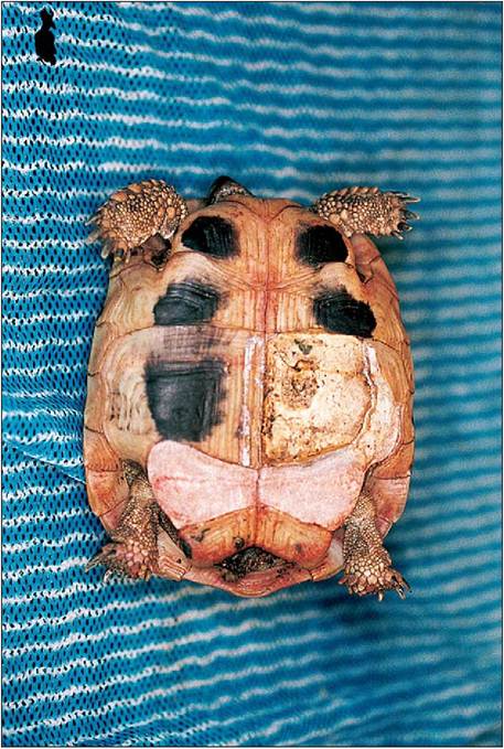
Figure 3.11 • Hermann’s tortoise (Testudo hermanni) with severe osteomyelitis and septicemia. The femoral and abdominal epidermal scutes have sloughed off to reveal the xiphiplastron and hypoplastron dermal bones beneath.
Pectoral girdle
The pectoral girdle consists of the epiplastron (clavicle), the entoplastron (interclavicle) and a tripartite arrangement of scapula, acromion process and coracoid bone. The scapula fuses dorsally with the carapace via a ligamentous attachment and ventrally articulates with the humerus at the glenoid cavity. A prominent acromion process projects medially, almost touching its counterpart and is fused to the plastron via connective tissue bands. The third strut is the coracoid bone, which extends caudomedially and also articulates with the glenoid fossa (Figs. 3.12 and 3.13) (Bellairs 1969a).
Limb girdles
The pectoral and pelvic girdle both occupy a unique position inside the ribs and act like vertical pillars giving extra strength to the shell. These two bony girdles are attached to the plastron and carapace by fan-shaped pectoral and pelvic muscles.
These limb muscles constitute the largest muscle mass and are surprisingly powerful. Fat is deposited between the limb base and the shell, so obese animals could have difficulties withdrawing into their shell and breathing.Pelvic girdle
The ilium, ischivin and pubic bones are paired and meet at the acetabulii; the ilium is attached dorsally to the sacral ribs. In Pleurodira the pelvic girdle is fused more strongly to the carapace by the ilia dorsally, and pubic and ischial bones ventrally, leaving the sacral attachment weaker (Hoffstetter & Gasc 1970).
Limbs
The humerus and femur are short in length, with expansion of the proximal and distal extremities. A fused carpus and
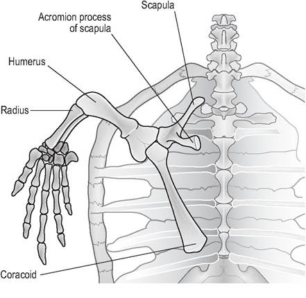
Figure 3.12 • Ventral view of tripartite chelonian shoulder. The scapula fuses dorsally with the carapace via a ligamentous attachment. A prominent acromion process projects medially while the coracoid bone extends caudomedially.
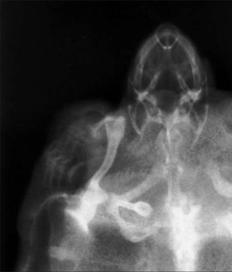
Figure 3.13 • Dorsal view of left shoulder of Red-eared slider (Trachemys scripta) showing tripartite shoulder.
tarsus all give added strength. All species have five claws on each foot, except the tortoises, which have short stubby toes and only four claws on the hind feet. The limbs are covered by conventional scales and have strong claws for digging. Freshwater turtles have webbed and flattened feet; the marine turtles have modified their forelimbs into flippers (Walker 1973).
Scutes
The outer part of the shell is covered with horny scutes formed from the epidermis and is the equivalent of scales in other reptiles. The number and size of scutes helps identify the species. The scutes, being of epidermal origin, are innervated and bleed if damaged. Growth occurs by the addition of new keratinized layers to the base of each scute. Greater cell activity around the perimeter of each new layer causes the scutes to widen gradually.
The scutes of the carapace are termed marginal, cervical, pleurals (costals) and vertebrals (Fig. 3.15). The scutes of the plastron are called gular, humeral, pectoral, abdominal, femoral, and anal (Fig. 3.16).
CLINICAL NOTE
It would be useful if chelonians could be aged by the ring of new growth in the shell, but as this can be interrupted by changes in food supply, seasonal change, and hibernation in temperate species it does not necessarily correspond with a year's growth (Enlow 1970; Hoffstetter & Gasc 1970).
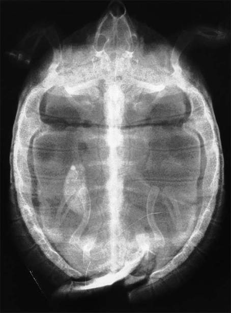
Figure 3.14 • Dorsoventral radiograph of carapace of juvenile Hermann's tortoise (Testudo hermanni) with shell fractured by lawn mower.
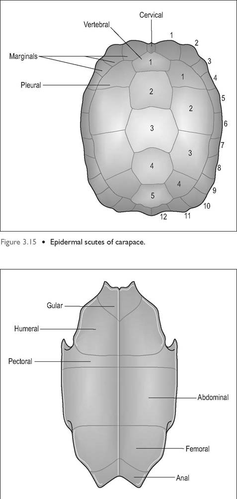
Figure 3.16 • Epidermal scutes of plastron.
Color and patterns
Many male Red-eared sliders (Trachemys scripta) go melanis- tic with age. When young they often have striking yellow plastrons, with clear patterns that become obliterated by pigment as they age (Hoffstetter & Gasc 1970).
Ecdysis
Like all reptiles, chelonians shed their skin but it tends to be in a piecemeal fashion. Aquatic terrapins shed their scutes as they grow, with the old scutes loosening first at the edges and then toward the center.
CLINICAL NOTE
Pyramidal growth of the shell may be caused by excess protein being available during the growing phase.
For example, if herbivorous tortoises are fed on dog food when juvenile this results in excessive growth surges and an imbalance in keratin production.
Shell modifications
Some species, like the Common box turtle (Terrapene carolina), have hinges of cartilaginous tissue between the sutures of the bony plates instead of the normal ossified sutures. These enable them to withdraw their head, tail, and limbs and close up like a box. The softshell turtles have leathery skin replacing hard scutes to make them more flexible. Marine species have reduced the bone plates even more, creating a tough, leathery shell for better streamlining. Other species, such as the Common snapping turtle (Chelydra serpintina), have such a small shell that they are unable to retract inside their shell at all. They escape predation by being extremely aggressive instead (Pough et al. 2002).
KEY POINTS
• Small head but incredibly strong retractor muscles
• Neck and tail are flexible but rest of spine is fused with shell, rib cage, and plastron
• No expandable rib cage
• Each shoulder is tripartite