Species Variations
5.2.1 Horse (Figure 5.1 and 5.2)
The duodenum is fairly firmly fixed by the mesoduodenum to the liver and the right dorsal colon. Immediately after the pylorus it turns sharply cranially through the sigmoid flexure and then caudally at the caudal flexure.
The remainder of the small intestine is very variable in position, lying in numerous coils with the small (descending) colon, chiefly in the left dorsal part of the abdomen from the stomach to the pelvic inlet.Initially the descending duodenum is related to the right kidney and the base of the caecum. It passes laterally and caudally around the base of the caecum and then crosses the midline. The ascending duodenum passes cranially on the left side, reaching the left kidney; there it continues into the jejunum. The small intestine of the horse is about 20 metres long.
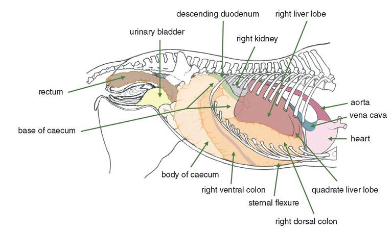
Figure 5.1 Right aspect of the gastrointestinal tract of a mare in situ.
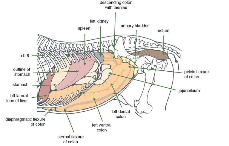
Figure 5.2 Left aspect of the gastrointestinal tract of the horse, in situ.
5.2.2 Ox (Figure 5.3)
The small intestine is up to 40 metres in length and 5-6 cm in diameter depending on the size of the animal. The duodenum is about 1 metre long and begins at the pylorus adjacent to the ventral end of the 10th rib on the right side. The diameter of the small intestine is 5-6 cm. The coils of the small intestine mostly occupy the ventral part of the right flank.
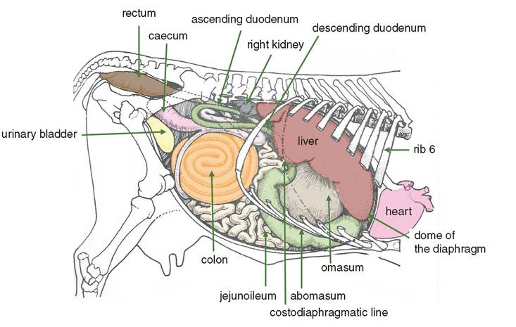
Figure 5.3
5.2.3 Sheep
The pylorus is located on the right side opposite the ventral end of the ninth rib. After passing dorsally and cranially to the visceral surface of the liver, the duodenum makes an S-shaped curve to reach the cranial pole of the right kidney.
The duodenum then passes caudally to the level of the tuber coxae, where it turns cranially at the iliac flexure and, at the level of the right kidney, becomes the jejunum. The coils of he jejunoileum reach the caecocolic junction on the medial side at the level of the last rib.5.2.4 Pig (Figure 5.4)
The stomach is fairly distended and contacts the body wall.
The duodenum is related to the liver and kidneys as in the dog. The jejunum lies mainly in the right caudal quadrant of the abdominal cavity but extends into the cau- doventral region on the left.
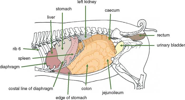
Figure 5.4 Left lateral aspect of the gastrointestinal tract of the pig, in situ.
5.2.5 Dog (Figure 5.5 and 5.6)
The small intestine of the dog is a narrow tube about two and a half times the length of the dog. The duodenum is relatively fixed in position by the short mesoduodenum. The cranial part of the duodenum is short and leaves the pylorus to lie across the liver from left to right; it is related to ribs 9 and 10 and lies in contact with the upper right flank. The descending duodenum passes caudally from the liver and on the ventral surface of the right kidney almost to the pelvic inlet. The ascending duodenum starts just cranial to the pelvic inlet and runs cranially on the left side of the abdomen, ventral to the descending colon and left kidney. The descending and ascending parts are in a V shape (Figure 2.1).
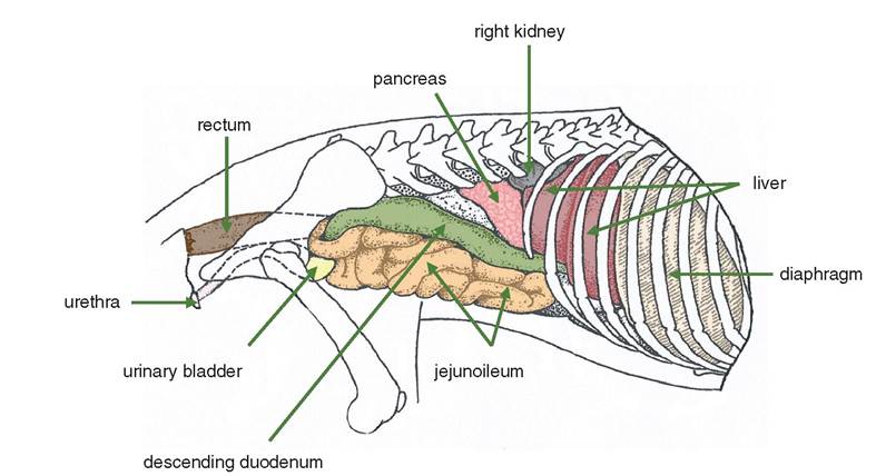
Figure 5.5 The right lateral view of the abdomen of the bitch.
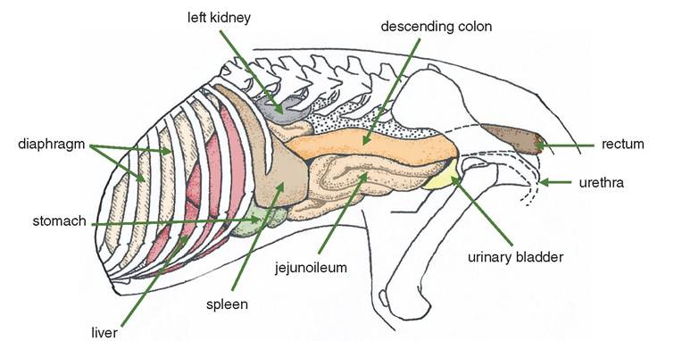
Figure 5.6 The left lateral view of the abdomen of the dog.
The jejunoileum is long and very mobile, in contrast to the duodenum and colon, which are short and relatively fixed in position. The jejunoileum is separated from the ventral and lateral body walls by the greater omentum (see Section 3.2).
5.3