Species Variations
6.2.1 Horse (Figures 5.1, 5.2 and 6.1)
The caecum resembles a gigantic comma, with the base dorsal in position and the body and the apex curving ventrally and cranially to reach the sternum.
Except for the apex, it lies to the right of the midline. The base is firmly attached to the right sublumbar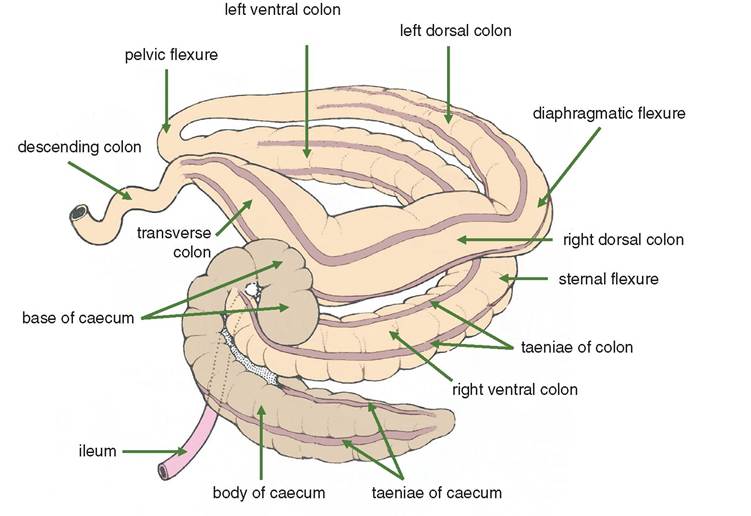
Figure 6.1 Caecum and ascending colon of the horse. The organs are viewed from the right side and from a slightly dorsal position. The body and apex of the caecum have been pulled ventrally and somewhat to the right.
region and to the right kidney. The descending duodenum passes around the base. The apex and much of the body lie on the abdominal floor, between the right ventral and left ventral colons. The caecum has four longitudinal muscle bands.
The large (ascending) colon has four divisions:
1) Right ventral colon: extends from the base of the caecum at about the ventral part of the last rib, along the costal arch and abdominal floor to the xiphoid cartilage. There it turns sharply as the sternal flexure. Four longitudinal muscle bands (taeniae coli) continue those of the caecum.
2) Left ventral colon: passes back along the abdominal floor, on the left of the caecum, to the general vicinity of the pelvic inlet. Here it bends sharply dorsally and cranially as the pelvic flexure. The pelvic flexure is very variable in its position and can cross to the right flank. There are again four longitudinal muscle bands. Beyond the pelvic flexure, the calibre of the colon is much reduced.
3) Left dorsal colon: much reduced in calibre, this runs cranially, lying dorsal or lateral to the left ventral colon, to reach the diaphragm and left lobe of the liver, and there turns back and to the right as the diaphragmatic flexure. Starting at the pelvic flexure, one longitudinal band is present, and this can be palpated per rectum.
4) Right dorsal colon: lies entirely within the thoracic cage. It passes caudally, dorsal to the right ventral colon. At the base of the caecum it turns medially, very close to the stomach and liver, and continues as the transverse colon. Three longitudinal muscle bands are present.
The transverse colon crosses from the right to the left side, cranial to the root of the mesentery. Its calibre is constricted, compared with the right dorsal colon.
The small colon (descending colon) begins at the end of the transverse colon, ventral to the left kidney and immediately caudal to the saccus caecus of the stomach. It mingles with the small intestine, occupying a similar position, i.e. mainly in the left dorsal part of the abdominal cavity. Its calibre is considerably less than that of the ascending colon. It has two longitudinal muscle bands and two rows of sacculations, and these features distinguish it from the small intestine.
6.2.2 Ox (Figures 4.5, 5.3 and 6.2)
The caecum extends caudodorsally in the upper part of the right flank. Its blind extremity terminates at the right side of the pelvic inlet, where it is related to the rectum dor- sally and the urinary bladder ventrally. The coils of the colon lie in the right caudal quadrant of the abdominal cavity. The general relations of the proximal loop, spiral loop and distal loop are shown in Figure 6.2.
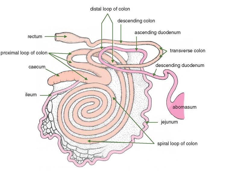
Figure 6.2 Semi-diagrammatic right lateral view of the intestines of the ox to show the proximal loop, the spiral loop and the distal loop of the ascending colon.
6.2.3 Sheep
The large intestine of the sheep is very similar to that of the ox. The caecum is about 10 inches long and 2 inches wide. The colon is about 15 feet long, and its diameter narrows distally.
6.2.4 Pig (Figures 5.4 and 6.3)
The caecocolic junction is dorsal and, on the left, related to the caudal pole of the left kidney.
The caecum is directed ventrally and caudally, covering the caudal aspect of the coils of the ascending colon. It lies in contact with the left abdominal wall. The apex often lies on the ventral body wall, more or less in the midline.The ascending colon forms a conical spiral. The direction taken by the axis of the spiral varies with the fullness of the stomach, but essentially it points ventrally and slightly to the left. The effect of this is to bring the spiral colon into extensive contact with the left abdominal wall as in Figure 6.3 The transverse colon lies in the usual position. The descending colon passes straight to the pelvic inlet close to the dorsal midline.
id="Picutre 27" class="lazyload" data-src="/files/uch_group31/uch_pgroup304/uch_uch7240/image/image027.jpg">
Figure 6.3 Diagram of the intestines of the pig as viewed from the right side.
6.2.2 Dog (Figures 5.5 and 5.6)
The caecum lies within the V of the duodenal loop, ventral to the right transverse processes of lumbar vertebrae two to four. It is related to the descending duodenum and the right kidney.
The ascending colon extends cranially on the right side of the abdominal roof, between the descending duodenum on its right and the small intestinal mass ventrally on its left. The transverse colon runs from right to left, cranial to the cranial mesenteric artery passing caudodorsally to the stomach. The descending colon lies against the upper part of the left flank covered by the greater omentum. It is related to the left kidney and the ascending duodenum.
6.3