Species Variations (Figure 12.2)
12.3.1 Horse
The kidneys of the horse are not ‘kidney-shaped' but are rather ‘heart-shaped'. The right kidney is compressed in a craniocaudal direction and is shorter than in the lateromedial dimension.
There is little or no external evidence of lobation. The renal pelvis is quite small, comprising only a centrally located collecting cavity. The renal crest is small, consisting only of the centrally located renal papillae. The papillary ducts at the extremities drain into one of two large collecting ducts called the terminal recesses, an arrangement found only in the horse.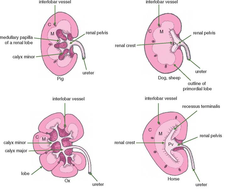
Figure 12.2 Diagrams showing the degree of Iobation of the mammalian kidney. Lobation is most marked in the largest mammals and in aquatic animals. C = cortex; M = medulla; shaded areas = renal sinuses occupied by fat and blood vessels. Pig: Lobes present but not visible externally. Several papillae drain into short calyces minores, which empty into two calyces majores. Dog and sheep: Single renal papilla elongated into a renal crest. Blood vessels show lobation. Ox: Lobes present and visible externally as well as internally. Each lobe has a papilla draining into a calyx minor, which empties into one or other of the two calyces majores. No renal pelvis. Horse: Lobation shown only by the blood vessels. The renal crest and the two recessi terminalis receive papillary ducts.
12.3.2 Ox
The right kidney of the ox is in contact with the caudate lobe of the liver. Both kidneys are surrounded by a large amount of fat termed the adipose capsule. The lobation of the embryonic kidney is retained in the ox, and there are about 20 lobes grossly visible in each kidney. Each lobe consists of a cortical bulge covering a medullary pyramid. The papillary ducts open into a common chamber called a renal pelvis that, in turn, opens into either the cranial or caudal branch of the ureter.
In adult ruminants the rumen causes the left kidney to be displaced to the right of the midline and to rotate so that the medial side faces dorsally.
12.3.3 Sheep
The kidneys of the sheep show no external sign of lobation so that the kidneys of this species are like those of the dog rather than the ox. The renal papillae are in a row called the renal crest that is surrounded by the renal pelvis.
12.3.4 Pig
Unlike the other domestic animals, the right kidney is not in contact with the liver. There is no external sign of lobation. Each renal papilla is surrounded by a minor calyx that opens into the renal pelvis or one of two major calyces at the extremities.
12.3.5 Dog/cat
In these species the anatomy of the kidneys is as described above in Sections 12.1 and 12.2. There is no external lobation, and the kidneys are bean-shaped and usually embedded in fat. The left kidney lies ventrally to the sublumbar muscles at the level of the second to fourth lumbar vertebrae; the right kidney lies adjacent to the first to third lumbar vertebrae and in a recess of the liver.
In most breeds the left kidney is palpable depending on the obesity of the individual. In the cat there are prominent capsular veins radiating from the hilus.
12.4