The Branches of the Abdominal Aorta
The listing and description of the major abdominal arteries given below is for the dog with added notes of important species variations. The coeliac, cranial mesenteric and caudal mesenteric arteries are single; all the other branches of the aorta are paired.
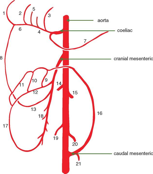
Figure 8.1 Ventral view diagram of the main arteries of the abdomen of the dog. 1 = right gastroepiploic; 2 = right gastric; 3 = left gastric; 4 = common hepatic; 5 = proper hepatic;
6 = gastroduodenal; 7 = splenic; 8 = cranial pancreaticoduodenal; 9 = middle colic; 10 = right colic; 11 = colic branch; 12 = common colic; 13 = caudal pancreaticoduodenal; 14 = right renal; 15 = left renal; 16 = left colic; 17 = ileocecocolic; 18 = jejunal; 19 = right testicular/ovarian; 20 = left testicular/ ovarian; 21 = cranial rectal.
8.1.1 Coeliac artery
This is the first visceral branch of the abdominal aorta. It is surrounded by a large plexus of nerves and the two coeliac ganglia (coeliacomesenteric plexus) together with numerous lymphatic vessels. The coeliac artery arises unpaired between the crura of the diaphragm and is only about 2 cm long. Usually it trifurcates into the common hepatic artery, left gastric artery and splenic artery. The common hepatic artery lies in a groove of the pancreas before it splits into several proper hepatic arteries supplying the liver parenchyma. The left of these hepatic arteries supplies the left lateral, left medial and quadrate lobes as well as a cystic branch to the gall bladder.
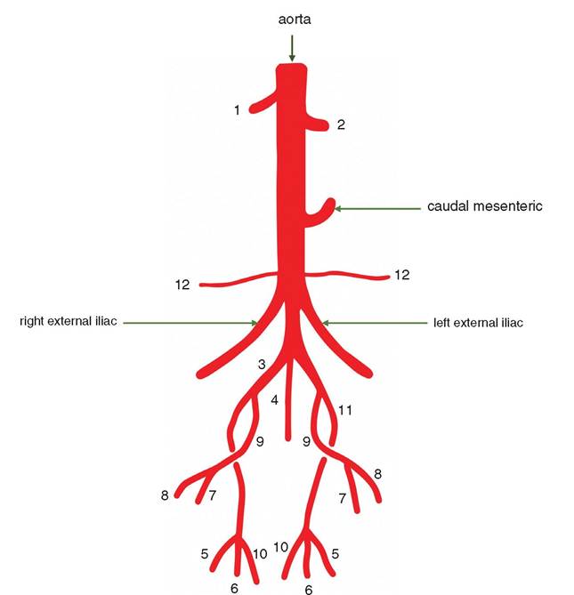
Figure 8.2 Diagram of the arteries of the pelvis of the dog. 1 = right testicular; 2 = left testicular/ ovarian; 3 = right internal iliac; 4 = median sacral; 5 = caudal gluteal; 6 = perineal; 7 = internal pududental; 8 = urogenital; 9 = visceral internal iliac; 10 = iliolumbar; 11 = parietal internal iliac; 12 = deep circumflex iliac.
The common hepatic artery becomes the gastroduodenal artery before terminating as the right gastroepiploic and cranial pancreaticoduodenal arteries. Both the numerous gastric and epiploic branches make numerous anastomoses in the stomach wall and the great omentum.
The splenic artery is a vessel of comparatively large diameter. It lies in the great omentum on its way to the spleen. It also has a branch to the pancreas and several small gastric branches that form anastomoses with branches of the left gastric artery.
8.1.2 Cranial mesenteric artery
This is the largest visceral branch of the abdominal aorta; it is about 5-10 mm caudal to the coeliac artery and about 5 mm in diameter; it is located at the level of the first or second lumbar vertebrae. Like the coeliac artery it is surrounded by the coeliacomesen- teric nerve plexus and numerous lymphatic vessels. It gives rise to several arteries
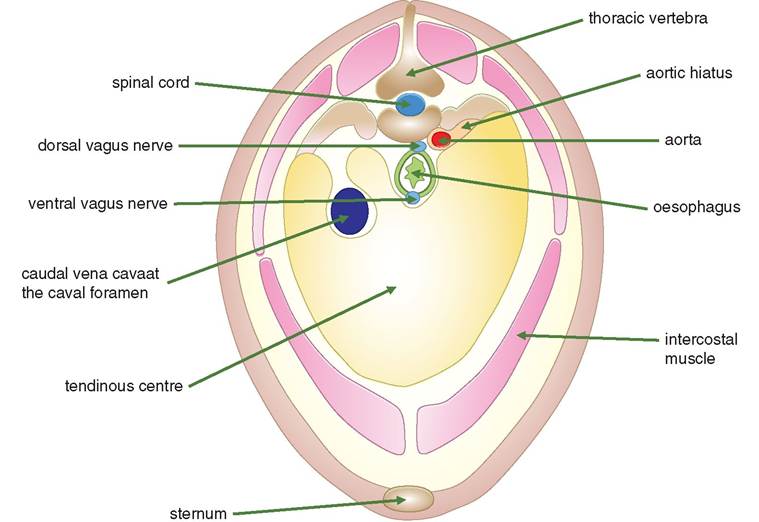
Figure 8.3 Diagram of the cranial view of the mammalian diaphragm. This example shows the azygos vein on the right so therefore must belong to a horse or a carnivore (see Section 9.1.2).
supplying blood to the intestines. Its branches are the common colic artery, the middle colic artery, the right colic artery, the ileocolic artery, the caudal pancreaticoduodenal artery and the multiple jejunal arteries.
The common colic artery is the first of the branches of the cranial mesenteric artery and is about 2 cm from the aorta. It gives rise to several branches, e.g. middle colic and right colic arteries, before becoming the ileocecocolic artery. There are several arterial branches of these vessels supplying the viscera and named according to the intestines and omenta that are supplied. In addition, there are many anastomoses.
8.1.3 Phrenicoabdominal arteries
These are paired parietal branches that supply the adrenal glands via the suprarenal arteries, the diaphragm via the caudal phrenic artery and the abdominal wall via the cranial abdominal artery.
It does seem likely that these two arteries are only found in carnivores. NAV is not clear on their existence in other species.8.1.4 Renal arteries
These are paired arteries supplying the kidneys. The right one is about 2 cm cranial to the left, conforming with the positioning of the kidneys, and about 3-4 mm in diameter. The renal arteries each supply at least two small branches to the caudal pole of the adjacent adrenal gland. The renal arteries often divide into several interlobar arteries before reaching the kidney hilus. See Chapter 12 for further discussion of the arterial supply to the kidneys.
8.1.2 Lumbar segmental arteries
The seven paired lumbar arteries are parietal branches of the aorta. Five of the arteries are branches of the abdominal aorta, and the cranial two are branches of the thoracic aorta. Each artery divides into a dorsal and a spinal branch. The dorsal branches supply the epaxial muscles and cutaneous tissues. The spinal branches enter the spinal canal with the spinal nerves.
8.1.3 Gonadal arteries
The paired ovarian arteries arise from the aorta about halfway between the renal and external iliac arteries. They follow a tortuous course through the broad ligaments to the ovaries. Branches supply the ovary, the ovarian bursa, the uterine cornu and the uterine tube. The branch to the uterine cornu anastomoses with the uterine artery, a branch of the internal iliac artery.
The testicular artery leaves the aorta at the corresponding level of the ovarian artery. Together with the testicular vein the artery enters the mesorchium (see Section 16.4 and Figure 16.2. This is the only artery supplying the testis and epididymis.
8.1.4 Caudal mesenteric artery
Arising as a single artery on the ventral aspect of the aorta at the level of the fifth lumbar vertebra in the dog (fourth lumbar vertebra in the horse), this artery reaches the distal extremity of the descending colon, where it bifurcates into the caudal rectal and left colic arteries; the latter anastomoses with the right colic artery.
The left colic artery lies within the mesocolon accompanying the descending colon until there is an anastomosis between the left colic and middle colic arteries.
8.1.5 Deep circumflex iliac arteries
These small paired arteries arise from the aorta about 1 cm cranial to the origin of the external iliac arteries. They supply the adjacent body wall via deep and superficial branches.
8.1.6 External iliac arteries
The external iliac artery is the largest parietal branch of the abdominal aorta. It is the principal artery to the hindlimb and divides into femoral and deep femoral branches before leaving the trunk to enter the hindlimb.
8.1.7 Internal iliac arteries
The aorta terminates just caudal to the origin of the external iliac arteries as a bifurcation into the pair of internal iliac arteries and the single median sacral artery.
The umbilical artery arises from the internal iliac near its origin and has different functions in the female depending on the presence of pregnancy. It is only functional during pregnancy when it carries blood from the aorta to the placenta. In the absence of pregnancy its lumen is obliterated, and it becomes the lateral umbilical ligament.
The internal iliac artery gives rise to visceral and parietal divisions. The urogenital and internal pudendal arteries belong to the visceral division. The parietal division includes the iliolumbar and the gluteal arteries.
In the male the artery of the penis terminates the internal pudendal artery and gives origin to the artery of the bulb of the penis, the deep artery of the penis and the dorsal artery of the penis.
In the female the cranial branch of the urogenital artery is the uterine artery. This artery varies in size depending on the physiological state of the reproductive tract. It courses through the broad ligament and sends small branches to the round ligament, the ovarian bursa and ovary. The artery of the clitoris is the terminus of the visceral internal pudendal artery.
8.2